Search results (1804 results)
-
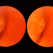
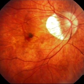
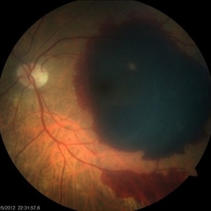
 Subhyaloid Hemorrhage
Oct 8 2012 by Jeffrey G. Gross, MD, FASRS
Subhyaloid hemorrhage, layered, with surrounding subretinal hemorrhage.
Condition/keywords: subhyaloid hemorrhage, subretinal hemorrhage
-
 Meridional Fold
Nov 9 2012 by Norman Byer
This is the same lesion as in the previous photograph. With the scleral indentation placed more posterior, we now can see that the fold ends over a small collection of subretinal fluid and that there is a very tiny retinal hole just below the posterior end of the retinal fold.
Condition/keywords: peripheral cystoid degeneration, retinal fold, retinal hole, scleral indentation, subretinal fluid
-
 White Retinal Tuft
Nov 9 2012 by Norman Byer
After six years, the previous lesion looked like this. The former flap has been completely avulsed and is now a free operculum. The white zone around the tear represents the small area of detachment and subretinal fluid. It is still asymptomatic and does not require treatment.
Condition/keywords: does not require treatment, free operculum, operculated retinal hole, subretinal fluid, white retinal tuft
-
 Subfoveal Subretinal Hemorrhage
Aug 28 2012 by Sharon Fekrat, MD FACS FASRS
subfoveal subretinal hemorrhage, right eye.
Photographer: Michael P. Kelly, FOPS Director, Duke Eye labs Duke University Eye Center Durham, NC
Imaging device: Zeiss FF40
Condition/keywords: subretinal hemorrhage
-
 Lattice Degeneration
Nov 9 2012 by Norman Byer
This 16-year-old girl has lattice degeneration and also this large oval retinal hole with a surrounding narrow zone of subretinal fluid. This lesion illustrates how large the atrophic holes of lattice degeneration may be. Occasionally the hole can be as large as the initial lattice lesion and can therefore obliterate all other evidence of its true identity. This was almost true in this case, but there does remain a small whitish remnant of the original lattice lesion at the lower end of the oval hole.
Condition/keywords: lattice degeneration, retinal hole, subretinal fluid, white lattice lines
-
 Inferior Rhegmatogenous Retinal Detachment with Subretinal Fibrosis
Aug 23 2012 by Gabriela Lopezcarasa Hernandez, MD
Asymptomatic 25-year-old woman with high myopia.
Photographer: Gabriela Lopezcarasa Hernandez, Hospital Angeles Lomas
Imaging device: FF4
Condition/keywords: high myopia, subretinal fibrosis
-
 ARMD with Disciform Scar
Oct 16 2012 by Jeffrey G. Gross, MD, FASRS
ARMD with disciform scar, RPE contracture, and subretinal hemorrhage, CF.
Condition/keywords: disciform scar, retinal pigment epithelium (RPE) contracture, subretinal hemorrhage
-
 Optos Giant Tear within Retinal Detachment
Apr 30 2019 by Lauren Whaley
Noticed an inferior visual field defect on a patient with history of vitreous hemorrhage. Decided to take an Optos image and this is what we found. Doctor performed pneumatic retinopexy in office and patient recovering well.
Photographer: Lauren R. Whaley
Imaging device: Optos
Condition/keywords: Optos, retinal tear, subretinal fluid
-
 Myopic CNV
Jan 11 2013 by Alex P. Hunyor, MD
Myopic macular degeneration complicated by subretinal neovascularisation, left eye.
Condition/keywords: high myopia, myopia, myopic choroidal neovascularization (CNV)
-
 Subretinal Hemorrhage
Sep 7 2012 by Raj K. Maturi, MD
Photographer: Char Harris, Midwest Eye Institute
Imaging device: TRC 50ex
-
 Myopic Choroidal Neovascular Membrane
Mar 25 2013 by Ratimir Lazic, MD, PhD
Color fundus photography of a 33-year-old female. In macular area subretinal hemorrhage can be seen. Area of atrophy temporal from PNO. Myopic changes of posterior pole and mid periphery can be noticed. The patient has been treated with 2 consecutive ranibizumab intravitreal injections. BCVA at baseline was 0,05 (Snellen lines) and 0,3 (Snellen lines) 2 months after.
Photographer: Marko Lukic, MD
Imaging device: Zeis Visucam Lite 2
Condition/keywords: high myopia, myopic choroidal neovascularization (CNV), ranibizumab
-
 Chronic Retinal Detachment: Features Slide 1
Oct 22 2012 by Ronald C. Gentile, MD
Chronic retinal detachments can be associated with demarcation lines (tidemarks), subretinal bands or sheets, and retinal cysts. Fundus photo of a chronic inferior retinal detachment reveals multiple demarcation lines inferior to the center of the fovea as a result of an inferior temporal dialysis.
Photographer: The New York Eye & Ear Infirmary Department of Medical Imaging
Condition/keywords: chronic retinal detachment, demarcation line
-
 Retinal Angiomatous Proliferation in Age-Related Macular Degeneration with Subretinal Neovascularization
Sep 24 2012 by James B. Soque, CRA, OCT-C, COA, FOPS
75-year-old white male with classic SRN with RAP. Lesion OD is active, and patient is receiving anti-VEGF treatment. Mid phase FA, 50 Deg, Mag 2x.
Photographer: James Soque, CRA, COA, Island Retina, Shirley, NY, USA
Imaging device: Topcon TRC 50 DX, OIS 5.0 MP Color, FA Camera, OIS Software
Condition/keywords: age-related macular degeneration (AMD), fundus autofluorescence (FAF), leakage, retinal angiomatous proliferation (RAP), subretinal neovascularization (SRNV)
-
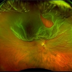
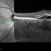 Recurrent Central Serous Choroidopathy
Aug 21 2012 by Edwin H. Ryan, MD
EDI-OCT showing thickened choroid and subretinal fluid
Photographer: Edwin Ryan Jr. MD, VitreoRetinal Surgery, PA
Imaging device: Heidelberg Spectralis
Condition/keywords: central serous chorioretinopathy (CSCR), choroidal thickening, enhanced depth imaging
-
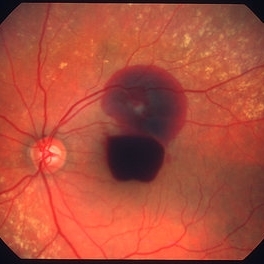 Ruptured retinal arterial macroaneurysm
Jan 11 2013 by Alex P. Hunyor, MD
Retinal arterial macroaneurysm with subretinal and preretinal hemorrhage
Condition/keywords: retinal arterial macroaneurysm
-
 Subhyaloid Hemorrhage
Oct 8 2012 by Jeffrey G. Gross, MD, FASRS
Subhyaloid hemorrhage, layered, with surrounding subretinal partial reabsorbed hemmorrhage, s/p YAG, hyaloidotomy.
Condition/keywords: hyaloidotomy, subhyaloid hemorrhage, subretinal hemorrhage
-
 Asymptomatic Lesion
Nov 9 2012 by Norman Byer
This asymptomatic lesion in a 27-year-old woman is a very interesting example of a cystic retinal tuft. Note the discrete white nubbin, which is the chief characteristic of this lesion. In this case, it is surrounded by a small area of subretinal fluid. The next slide pair will reveal the reason for this.
Condition/keywords: asymptomatic, cystic retinal tuft, subretinal fluid
-
---thumb.JPG/image-square;max$300,300.ImageHandler) Retinal Pigment Epithelial Detachment With No Subretinal Fluid
Retinal Pigment Epithelial Detachment With No Subretinal Fluid
Jun 29 2013 by Jason S. Calhoun
A 38-year-old male who comes in with blurred vision in the left eye. VA is 20/30. Noticed a defect inferior of his central vision. Did an fluorescein angiogram to determine an RPE with no sub retinal fluid. Also OCT confirms. Patient was injected with Avastin.
Photographer: Jason S. Calhoun, Mayo Clinic Jacksonville, Florida
Imaging device: TOPCON TRC 50-EX
Condition/keywords: central serous retinopathy (CSR), retinal pigment epithelium (RPE) detachment
-
 Choroidal Rupture with Subretinal Hemorrhage
Oct 1 2012 by Jeffrey G. Gross, MD, FASRS
Choroidal rupture with subretinal hemorrhage.
Condition/keywords: choroidal rupture, subretinal hemorrhage
-
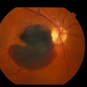
 Choroidal Melanoma Large Amelanotic
Oct 15 2012 by Susanna S. Park, MD, PhD
68-year-old man with a large amelanotic mass and subretinal fluid in the left eye. Visual acuity was CF and extrascleral extension was suspected on MRI scan.
Photographer: Ellen Redenbo, University of California Davis Eye Center
Condition/keywords: melanoma
-
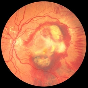
 Massive Submacular Hemorrhage
Sep 28 2012 by Joseph M. Civantos, MD
82-year-old gentleman who developed this massive submacular hemorrhage 3 days after his 16th Lucentis injection. Visual acuity dropped from 20/80 to LP.
Condition/keywords: subretinal hemorrhage
-
 AMD with Subretinal Hemorrhage Recurrence OS
Aug 24 2012 by John S. King, MD
Six months after subretinal tPA and regular antiVEGF, last of which was Eylea, there was a recurrent hemorrhage, and acuity drop from 20/50 to HM; discussed repeat subretinal tPA.
Photographer: Kristin Konecki, OcuSight Eye Care Center, Rochester, NY
Condition/keywords: EYLEA, subretinal hemorrhage
-
 Secondary Choroidal Neovascularization Due to Toxoplasmosis
Feb 25 2013 by Henry J. Kaplan, MD
Left eye: secondary choroidal neovascularization and subretinal hemorrhage in a patient with old macular scar of toxoplasma.
Condition/keywords: choroidal neovascularization (CNV), toxoplasmosis, toxoplasmosis chorioretinitis
-
 White Retinal Tuft
Nov 9 2012 by Norman Byer
This white retinal tuft was seen in a 20-year-old man. It is associated with an asymptomatic retinal tear posterior to the tuft and with a tiny adjacent amount of subretinal fluid. It remained just like this for six years and then underwent the change shown in the next slide pair.
Condition/keywords: asymptomatic, subretinal fluid, white retinal tuft
-
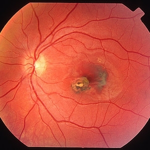
 Chronic Macular Hole
Sep 2 2012 by Hyung-Woo Kwak, MD
A large hole with rolled everted edges, adjacent cystoid intraretinal spaces, a shallow rim of subretinal fluids.
Imaging device: Zeiss F450 plus
Condition/keywords: macular hole
 Loading…