Search results (1804 results)
-
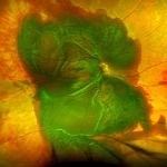
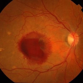 Macroaneurysm
Apr 1 2017 by Manish Nagpal, MD, FRCS (UK), FASRS
Case of a ruptured macroaneurysm with subhyaloid and subretinal blood.
Photographer: Avijit Vishnoi
Condition/keywords: macroaneurysm, ruptured macroaneurysm
-
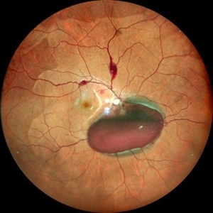
 Massive Subretinal Hemorrhage With Near Total Retina Detachment
Nov 27 2013 by David W. Faber, MD
Fundus photo of an 71-year-old male with massive subretinal hemorrhage. Had been given 6 week Avastin treatments. Was put on coumadin for 6 weeks following knee surgery.
Photographer: Donna Knight, Rocky Mountain Retina Consultants, Salt Lake City, Utah
-
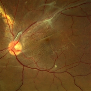
 Choroidal Melanoma
Nov 3 2022 by pedro fernandes souza neto
Transillumination of Enucleation specimen of Choroidal Melanoma: anterior chamber is closed. Total secondary retinal detachment with subretinal serous fluid and some subretinal hemorrhages are present.
Photographer: Eduardo Marback, Federal University of Bahia, Brazil
Condition/keywords: enucleation, melanoma
-
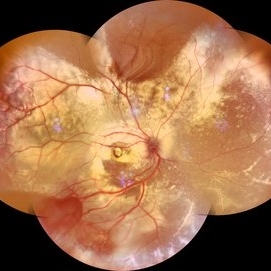
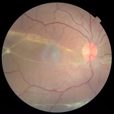
 Valsalva Retinopathy
Dec 20 2021 by Unnati Vishwanath Shukla, M. S. ,DNB, FVRS FNERF, MNAMS,PhD Scholar(Retina)
26-year-old male with Valsalva Retinopathy. History of severe cough for 3 days. All hematological investigations were within normal limits.
Photographer: Dr. Unnati Shukla, Consultant, Retina Foundation, Ahmedabad
Imaging device: Nidek Mirante
Condition/keywords: subhyaloid hemorrhage, subretinal hemorrhage, valsalva retinopathy
-
 Disciform Scar
Aug 18 2020 by Aditya S Kelkar, MS, FRCS, FASRS,FRCOphth
Left eye fundus photograph of 75-year-old male, showing large disciform scar post subretinal bleeding secondary to idiopathic polypoidal choroidal vasculopathy
Photographer: Dr.Mounika Bolisetty
Imaging device: CLARUS 500
Condition/keywords: disciform scar, idiopathic polypoidal choroidal vasculopathy
-
 Displaced & folded macula
Oct 10 2022 by Ricardo Leitão Guerra
Tractional retinal detachment due to sickle cell retinopathy leading to a displaced and folded appearance of the macula in this 36-yo male. Subretinal bands are also noticed crossing the macula towards inferior retinal detachment area.
Photographer: Ricardo Leitão Guerra
Imaging device: Clarus 700 - Zeiss
Condition/keywords: folds, sickle cell retinopathy, subretinal bands, tractional retinal detachment
-
 Lady in a dress
Feb 9 2023 by Shelby Helton
Fluorescein Angiography on a 67-year-old male with significant RPE changes secondary to a severe subretinal hemorrhage that required a vitrectomy with subretinal TPA in 2013.
Photographer: Shelby Helton
Imaging device: Heidelberg Spectralis
Condition/keywords: wet age-related macular degeneration (wet AMD)
-
 Subretinal BSS and air
Apr 12 2022 by Nassim Alejandro Abreu Arbaje, MD
67 year old female who presented with complaints of 5 days of decreased vision of her left eye. She underwent PPV + BSS and Air injection in the subretinal space
Photographer: Nassim Abreu, Dr. Elias Santana Hospital
Imaging device: Ngenuity 3D system screenshot
Condition/keywords: subretinal hemorrhage
-
 Central Serous Retinopathy
Mar 19 2024 by Corey Grant
Ultra Wide-Field Fundus Autofluorescence Imaging of a 37 year old female with Central Serous Retinopathy affecting her right eye. Patient Visual Acuity was 20/20 in both eyes. Patient reported black spots in her vision onset three years ago, with associating flashes of light. Patient reports history of cortisone back injections a few years ago and denies Flonase use. The physician stated that there is hyperautofluorescence in the area of gutter of Sub-Retinal Fluid which likely happened from CSR.
Photographer: Corey Grant, OSC
Imaging device: OPTOS CALIFORNIA RGB
Condition/keywords: Central Serous Chorioretinopathy (CSR), central serous retinopathy (CSR), fundus autofluorescence (FAF), Guttering, hyperautofluorescence, inferior retina, OPTOS, Retina, Right Eye, subretinal fluid, ULTRA WIDE FIELD
-
 Coats' Disease Montage
Feb 5 2021 by Akansha Sharma
Fundus photograph of a 5-year-old male child who presented with unilateral diminution of vision since one month.
Photographer: Dr. Nivesh Gupta, M.S., Retina Foundation, Ahmedabad
Condition/keywords: angiomatosis retinae, Coats' disease, exudative detachment, subretinal exudates
-
 Idiopathic Uveal Effusion Syndrome
Aug 22 2024 by Jordyn Beckman
61 year old male with Idiopathic Uveal Effusion Syndrome with starry night appearance on fluorescein. 3 weeks s/p single external drainage retinotomy and 9 weeks of oral pred with recurrent choroidal effusions. Has since returned to surgery for secondary drainage retinotomy; subretinal fluid remain persistent.
Photographer: Jordyn Beckman
Imaging device: Optos California
Condition/keywords: chorioretinitis, Choroidal, exudative detachment, window defect
-
 Massive SRH in Patient on Coumadin Being Treated for Exudative AMD
Sep 30 2019 by John S. King, MD
78-year-old white female using 1mg of warfarin for atrial fibrillation, who had a large PED, Type 1 lesion from AMD. Noticed acute darkening of vision one week after anti-VEGF injection. Has very large SRH, subRPE heme, and corrugated retinal appearance post RPE tear. Vision HM (from 20/100). 20/25 in the fellow eye that has dry AMD.
Photographer: Shelly Blair
Imaging device: Optos CA
Condition/keywords: subretinal hemorrhage, wet age-related macular degeneration (wet AMD)
-
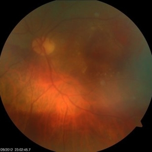
 Massive Submacular Hemorrhage
Sep 28 2012 by Joseph M. Civantos, MD
82-year-old gentleman who developed this massive submacular hemorrhage 3 days after his 16th Lucentis injection. Visual acuity dropped from 20/80 to LP.
Condition/keywords: subretinal hemorrhage
-
 Optic Nerve Melanocytoma
Apr 3 2023 by Gustavo Aguirre Suarez
Fundus photograph of a 36-year-old female with a lesion dependent on the optic nerve head with subretinal extension, elevated, about 1.5 disc diameters, dark brown to black in color, involving more than three quarters of the neuroretinal ring towards the inferonasal area.
Photographer: Dr. Gustavo Aguirre-Suarez
Imaging device: Zeiss Visucam 500
Condition/keywords: melanocytic lesion, Melanocytoma
-
 PEHCR (Peripheral Exudative Hemorrhagic Chorioretinopathy)
May 12 2023 by Niloofar Piri, MD
Ultrawide fundus photograph of the left eye demonstrating extensive peripheral hemorrhagic exudative detachment in a 79 yo Caucasian female with prior history of non-exudative AMD. Recent diagnosis of Acute myeloid leukemia with low platelet count which might have contributed to the above presentatuon. Please note the temporal subretinal hemorrhage as well as RPE atrophy and hyperplasia in the macula.
Photographer: Rocio Bentivegna, MD, Saint Louis University; Jessica Maddox, COA, Saint Louis University
Condition/keywords: peripheral exudative hemorrhagic chorioretinopathy (PEHCR)
-
 Post Subretinal tpa , viterectomy and gas
May 6 2022 by Shobhit Chawla, M.S.
SUBMACULAR HAEMORRHAGE IN A 38YEAR OLD LADY PATIENT CAUSE POLYP BLEED IN PCV. Following viterectomy , subretinal tpa . gas and aflibercept injection. 7 day post operative image.
Photographer: Shobhit Chawla
Imaging device: Zeiss Clarus 500
Condition/keywords: aflibercept, intravitreal gas bubble, submacular hemorrhage, tissue plasminogen activator (tPA), vitrectomy
-
 Retinal Detachment With Subretinal Cords
Apr 30 2015 by Mitzy E Torres Soriano, MD
Retinal detachment with sub retinal cords.
Photographer: Mitzy E. Torres Soriano, MD; Centro medico Cagua-Estado Aragua. Venezuela
Imaging device: TOPCON
Condition/keywords: proliferative vitreoretinopathy (PVR), subretinal cords
-
 Sunset Glow Fundus
May 15 2022 by Manuel Ángel Alcántara Delgado, MD
Optomap ultra-widefield retinal imaging of an 35-year-old woman showed sunset glow fundus, multiple nummular chorioretinal atrophic lesions, macular subretinal fibrosis and pigment clumping in chronic recurrent stage of Vogt-Koyanagi-Harada disease.
Photographer: Manuel Ángel Alcántara Delgado. Conde de Valenciana.
Condition/keywords: abnormal retina, benign pigmented lesions, pigment clumps, retinal fibrosis, uveitis, Vogt-Koyanagi-Harada
-
---thumb.jpg/image-square;max$300,300.ImageHandler) Active Choroidal Neovascularization With Subretinal Hemorrhage
Active Choroidal Neovascularization With Subretinal Hemorrhage
Nov 25 2013 by Maurice F. Rabb
Active choroidal neovascularization with subretinal hemorrhage.
Condition/keywords: choroidal neovascularization (CNV), subretinal hemorrhage
-
 AMD with Subretinal Hemorrhage OS
Aug 24 2012 by John S. King, MD
One week s/p subretinal tPA; acuity improved from HM to 20/100.
Photographer: Kristin Konecki, OcuSight Eye Care Center, Rochester, NY
Condition/keywords: subretinal hemorrhage
-
 Amelanotic Choroidal Melanoma
May 18 2020 by McGill University Health Centre
The enucleation specimen in (A) shows an amelanotic, mushroom-shaped tumor arising from the choroid. Microhemorrhages are present within the tumor and also surround the tumor base (arrow). True retinal detachment is present (arrowhead). The subretinal fluid is mixed: clear (1), hemorrhagic (2), and fibrinoid (3).
Condition/keywords: enucleation, mushroom-shaped
-
 Amelanotic Mushroom-Shaped Choroidal Melanoma
May 18 2020 by McGill University Health Centre
This enucleation specimen demonstrates an amelanotic, mushroom-shaped, slightly hemorrhagic tumor near the optic nerve (arrow). True retinal detachment is present, and the retina is folded (arrowhead). The subretinal fluid is hazy (*).
Condition/keywords: amelanotic melanoma, enucleation, mushroom-shaped
-
 Branch Retinal Vein Occlusion
Oct 17 2012 by Sharon Fekrat, MD FACS FASRS
Fluorescein angiography of an inferior perfused branch retinal vein occlusion in the left eye of a middle aged male with hypertension. The foveal avascular zone is irregular. Subretinal hemorrhage is present.
Photographer: John Reaves, Ophthalmic Photographer, Durham VA Medical Center Eye Clinic Imaging Suite, Durham, NC
Imaging device: Fluorescein Angiography
Condition/keywords: branch retinal vein occlusion (BRVO), subretinal hemorrhage
-
 Bullous Retinal Detachment
Jul 9 2021 by Anton Orlin, MD
This is a color photograph of a right eye with a superior, bullous, macula-splitting retinal detachment. These features place the patient at a higher risk for macular fold formation postoperatively. To prevent fold formation, a surgeon should attempt for more complete subretinal fluid drainage during repair. This can be done with the use of perfluorocarbon liquid or by making a drainage retinotomy.
Condition/keywords: macular fold
-
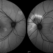 Central Serous Chorioretinopathy
Jan 25 2022 by Olivia Rainey
Widefield fundus autofluorescence of a 60-year-old male with Central Serous Chorioretinopathy affecting both eyes. Chronic history of CSR followed with observation without treatment prior to presenting at our office. The physician noted significant findings on exam and imaging with multifocal areas of inactive and active changes in the right eye and subfoveal subretinal fluid with recent visual decline in the left eye. There are hyper and hypoautofluorescent changes, consistent with CSR.
Photographer: Olivia Rainey, OCT-C, COA
Imaging device: Heidelberg Spectralis
Condition/keywords: 55-degrees, central serous chorioretinopathy (CSCR), central serous retinopathy (CSR), chronic central serous chorioretinopathy (CSCR), fundus autofluorescence (FAF), heidelberg spectralis, left eye
 Loading…