 Initializing download.
-
 By Norman Byer
From Dr. Norman E. Byer’s “The Peripheral Retina in Profile” - Uploaded on Nov 9, 2012.
- Last modified by Suber S. Huang, MD, MBA, FASRS on Feb 10, 2013.
- Reviewed by Chayal Patel
- Rating
- Appears in
- Miscellaneous
- Condition/keywords
- lattice degeneration, retinal hole, subretinal fluid, white lattice lines
- Description
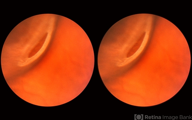
- This 16-year-old girl has lattice degeneration and also this large oval retinal hole with a surrounding narrow zone of subretinal fluid. This lesion illustrates how large the atrophic holes of lattice degeneration may be. Occasionally the hole can be as large as the initial lattice lesion and can therefore obliterate all other evidence of its true identity. This was almost true in this case, but there does remain a small whitish remnant of the original lattice lesion at the lower end of the oval hole.


---thumb.JPG/image-square;max$79,0.ImageHandler "Central Serous Retinopathy with Fibrin - Mid-phase FA")

---thumb.JPG/image-square;max$79,0.ImageHandler "Central Serous Retinopathy with Fibrin")
---thumb.jpg/image-square;max$79,0.ImageHandler "Lattice Degeneration")
