 Initializing download.
-
 By Ratimir Lazic, MD, PhD
Eye Clinic Svjetlost
Co-author(s): Marko Lukic, MD - Uploaded on Mar 25, 2013.
- Last modified by Ratimir Lazic, MD, PhD on Mar 25, 2013.
- Reviewed by Ratimir Lazic, MD, PhD
- Rating
- Appears in
- Myopic CNV
- Condition/keywords
- high myopia, myopic choroidal neovascularization (CNV), ranibizumab
- Photographer
- Marko Lukic, MD
- Imaging device
-
Fundus camera
Zeis Visucam Lite 2 - Description
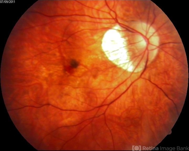
- Color fundus photography of a 33-year-old female. In macular area subretinal hemorrhage can be seen. Area of atrophy temporal from PNO. Myopic changes of posterior pole and mid periphery can be noticed. The patient has been treated with 2 consecutive ranibizumab intravitreal injections. BCVA at baseline was 0,05 (Snellen lines) and 0,3 (Snellen lines) 2 months after.


