Search results (2916 results)
-
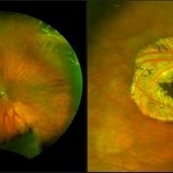
 Cystic Retinal Tuft
Nov 9 2012 by Norman Byer
This is the same lesion as in the previous slide pair but the photograph was taken nine years later when the patient was 58-years-old soon after an acute posterior vitreous detachment. This demonstrates that posterior vitreous detachment can produce large retinal tears at these sites. However, it is important to emphasize that prophylactic treatment of cystic retinal tufts in the absence of a retinal tear would be very ill-advised because several hundred innocence and harmless lesions would have to be treated in order to prevent one tear of the retina.
Condition/keywords: cystic retinal tuft, posterior vitreous detachment, retinal tear
-
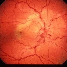
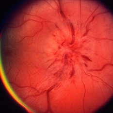 Papillitis
May 2 2013 by Henry J. Kaplan, MD
Anterior optic neuropathy or papillitis in the right eye; notice the blurred optic disc margin, engorged capillaries and flame shaped hemorrhages at the margin.
Condition/keywords: optic disc edema, optic disc swelling, papillitis
-
 Lattice Degeneration
Nov 9 2012 by Norman Byer
This is a more typical classical example of lattice degeneration in a 42-year-old woman in a photograph taken without scleral indentation. It shows much more marked vascular changes than the previous case. Note the tapering of the blood columns as the vessels approach the lesion and also the white sheathing of the vessel walls. Note also the continuity of the blood vessels on opposite sides of the lesion with the characteristic white lattice lines. More than 45 years ago Vogt pointed this out as a proof that these white lines were actually caused by changed blood vessels. Note also that this lesion shows a combination of several individual features of lattice degeneration. In addition to the white lines, there is a reddish crater-like area beneath the main horizontal white line. There is a prominent horizontal zone below this white line showing a snailtrack appearance. Also, there are two tiny atrophic retinal holes outside the photograph on the right end of this lesion. This eye contained five such retinal holes and they have all remained unchanged for more than 10 years of observation without treatment.
Condition/keywords: atrophic retinal hole, lattice degeneration, moderate snail track, tapering blood columns, white lattice lines, white sheath vessel
-
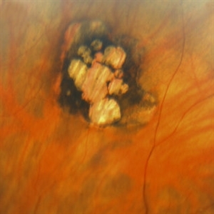
 Congenital Hypertrophy of the Retinal Pigment Epithelium (CHRPE)
Mar 1 2014 by Homayoun Tabandeh, MD, FASRS
Congenital hypertrophy of the retinal pigment epithelium (CHRPE).
Condition/keywords: congenital hypertrophy of the retinal pigment epithelium (CHRPE)
-
---thumb.jpg/image-square;max$300,300.ImageHandler) Presumed Ocular Histoplasmosis Syndrome
Presumed Ocular Histoplasmosis Syndrome
Feb 26 2013 by Henry J. Kaplan, MD
Color fundus photograph of the right eye of a patient with POHS shows typical punched out scars and peripapillary atrophy.
Condition/keywords: presumed ocular histoplasmosis syndrome (POHS)
-
---thumb.jpg/image-square;max$300,300.ImageHandler) Myopic fundus
Myopic fundus
Jan 11 2013 by Hyung-Woo Kwak, MD
Myopic fundus reveals yellow-colored lacquer cracks and peripapillary atrophy. There was visible choroidal vessel due to thin retina.
Photographer: Misook Lee, Kyung Hee Univsersity Hospital, Seoul
Imaging device: Zeiss f 450 plus
Condition/keywords: myopic fundus
-
 Lattice Degeneration
Nov 9 2012 by Norman Byer
This 16-year-old girl has lattice degeneration and also this large oval retinal hole with a surrounding narrow zone of subretinal fluid. This lesion illustrates how large the atrophic holes of lattice degeneration may be. Occasionally the hole can be as large as the initial lattice lesion and can therefore obliterate all other evidence of its true identity. This was almost true in this case, but there does remain a small whitish remnant of the original lattice lesion at the lower end of the oval hole.
Condition/keywords: lattice degeneration, retinal hole, subretinal fluid, white lattice lines
-
---thumb.jpg/image-square;max$300,300.ImageHandler) Geographic atrophy
Geographic atrophy
Aug 29 2012 by Young Hee Yoon, MD, PhD
OCT image of an 78-year-old woman. Her best-corrected visual acuity was counting fingers at 30cm.
Photographer: Ji Hee Kim, Asan Medical Center
Imaging device: Heidelberg spectralis
Condition/keywords: dry age-related macular degeneration (dry AMD), geographic atrophy
-
---thumb.jpg/image-square;max$300,300.ImageHandler) Inferior Sector Iris Atrophy With Depigmentation
Inferior Sector Iris Atrophy With Depigmentation
Aug 1 2013 by From the Collections of Thomas M. Aaberg, MD and Thomas M. Aaberg Jr., MD
Inferior sector iris atrophy with depigmentation.
Condition/keywords: depigmentation, inferior sector iris atrophy
-
 Geographic Atrophy, Fundus photograph
Aug 23 2012 by Gerardo Garcia-Aguirre, MD
Fundus photograph of an 85-year-old patient with age related macular degeneration and geographic atrophy. A large area with well-defined borders is observed, in which the choroidal vasculature is visualized.
Photographer: Noemí Hernández, Asociación para Evitar la Ceguera en México
Imaging device: Zeiss FF4
Condition/keywords: geographic atrophy
-
 Stage 5 Retinopathy of Prematurity (ROP)
Oct 9 2012 by Audina M. Berrocal, MD FASRS
Advanced APROP with Stage 5 and vascularly active.
Photographer: Ditte Hess CRA, BPEI
Imaging device: RETCAM
Condition/keywords: retinopathy of prematurity (ROP), stage 5
-
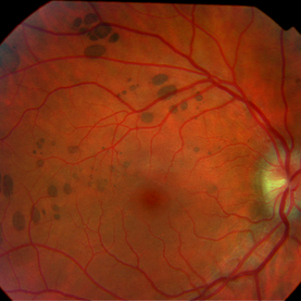 Bear Tracks
Dec 31 2012 by Raj K. Maturi, MD
Photographer: Tom Steele, CRA Midwest Eye Institute Indianapolis, Indiana
Imaging device: Topcon 50ex 50 degree field
Condition/keywords: bear tracks, benign pigmented lesions, congenital hypertrophy of the retinal pigment epithelium (CHRPE), OD
-
 Myopic Choroidal Neovascular Membrane
Mar 25 2013 by Ratimir Lazic, MD, PhD
Color fundus photography of a 33-year-old female. In macular area subretinal hemorrhage can be seen. Area of atrophy temporal from PNO. Myopic changes of posterior pole and mid periphery can be noticed. The patient has been treated with 2 consecutive ranibizumab intravitreal injections. BCVA at baseline was 0,05 (Snellen lines) and 0,3 (Snellen lines) 2 months after.
Photographer: Marko Lukic, MD
Imaging device: Zeis Visucam Lite 2
Condition/keywords: high myopia, myopic choroidal neovascularization (CNV), ranibizumab
-
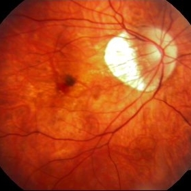
 Central Retinal Artery Occlusion & Cilioretinal Artery Sparing
Dec 22 2012 by Hamid Ahmadieh, MD
Early phase FA image of the right eye of a 34-year-old man with sudden drop of vision due to CRAO. The macula is involved despite cilioretinal artery sparing .
Photographer: Zohre Salimi; Labbafinejad Medical Center, Shahid Beheshti University of Medical Sciences , Tehran
Imaging device: Heidelberg HRA
Condition/keywords: central retinal artery occlusion (CRAO), cilioretinal sparing
-
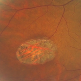
 Atrophic Holes in Lattice Lesion
Nov 9 2012 by Norman Byer
In this 26-year-old woman, these two atrophic holes in a lattice lesion led to a clinical retinal detachment which was operated on successfully. In retinal detachments of this type resulting from non tractional atrophic holes, it has been found that 50% occur before the age of 30 years.
Condition/keywords: atrophic retinal hole, lattice lesion
-
---thumb.jpg/image-square;max$300,300.ImageHandler) Reticular Pattern Dystrophy
Reticular Pattern Dystrophy
Aug 7 2013 by From the Collections of Thomas M. Aaberg, MD and Thomas M. Aaberg Jr., MD
Color fundus photograph reveals typical reticular - type pattern dystrophy, OS.
Condition/keywords: pattern macular dystrophy, reticular dystrophy
-
 Operculated Hole and CHRPE
Jan 16 2018 by Carolyn Daley
58-year-old woman with an operculated hole and CHRPE in the right eye. Patient is asymptomatic so no treatment was recommended at this time.
Photographer: Carolyn Daley
Imaging device: Optos ultra wide field image
Condition/keywords: congenital hypertrophy of the retinal pigment epithelium (CHRPE), operculated retinal hole, Optos, ultra-wide field imaging
-
 Sudden Posterior Vitreous Detachment
Nov 9 2012 by Norman Byer
This is the appearance of the previous lesion three weeks following prophylactic cryotherapy. Continuing vitreal retinal traction has a now torn the flap completely free from the retina. The whitish cystic retinal tuft can be discerned on the upper part of the free operculum. Along the lower half of the operculum superimposed over the dark shadow of the scleral indentation one may observe numerous, delicate, vitreous fibrils actually attaching to the operculum.
Condition/keywords: cystic retinal tuft, free operculum, prophylactic cyrotherapy, retinal flap, scleral indentation, vitreoretinal traction, vitreous fibrils
-
 AION With Ciliotretinal Artery Occlusion
May 2 2013 by Henry J. Kaplan, MD
AION accompanied by partial CRAO which is visible as retinal edema and cherry red spot.
Condition/keywords: anterior ischemic optic neuropathy, central retinal artery occlusion (CRAO)
-
 Asymptomatic Rhegmatogenous Retinal Detachment
Sep 14 2012 by Sharon Fekrat, MD FACS FASRS
Fundus photograph of a 25-year-old emmetropic male graduate student with an inferotemporal phakic chronic asymptomatic rhegmatogenous retinal detachment with a demarcation line in the right eye. His sister who is an ophthalmology resident discovered this incidental finding. Vision 20/20.
Photographer: Brian Lutman CRA, Duke University Eye Center, Durham, NC
Condition/keywords: asymptomatic, demarcation line
-
 Rhegmatogenous Retinal Detachment in Retinopathy of Prematurity
Oct 9 2012 by Audina M. Berrocal, MD FASRS
45-week-old ex-premature 24-week child who had a rhegmatogenous detachment after laser
Photographer: Ditte Hess CRA, BPEI
Imaging device: Ret Cam
Condition/keywords: laser, retinopathy of prematurity (ROP)
-
 Stargardt macular dystrophy slide 1
Oct 22 2012 by Ronald C. Gentile, MD
16-year-boy with difficulty in school seeing the black board. The macula area of the right eye had areas with a beaten bronze appearance and atrophy. Small pisci-form flecks can be seen surrounding the fovea.
Photographer: The New York Eye & Ear Infirmary Department of Medical Imaging
Condition/keywords: small pisci-form flecks, Stargardt disease
-
 Acute Idiopathic Occlusive Retinal Vasculitis
May 31 2014 by Hamid Ahmadieh, MD
Color fundus photograph of the right eye of a 28-year-old woman with sudden drop of vision due to acute occlusive retinal vasculitis leading to extensive nerve fiber layer infarction and retinal hemorrhages.
Photographer: Naghmeh Nozhat, Negah Eye Center, Tehran
Condition/keywords: color fundus photograph, cotton wool spots, retinal hemorrhage, retinal ischemia
-
---thumb.jpg/image-square;max$300,300.ImageHandler) Anterior Ischemic Optic Neuropathy
Anterior Ischemic Optic Neuropathy
Mar 29 2013 by Henry J. Kaplan, MD
Anterior Ischemic Optic Neuropathy; notice the typical pale optic disc swelling and faint splinter hemorrhages.
Condition/keywords: anterior ischemic optic neuropathy
-
 Congenital Hypertrophy of the Retinal Pigment Epithelium (CHRPE)
Mar 1 2014 by Homayoun Tabandeh, MD, FASRS
Congenital hypertrophy of the retinal pigment epithelium (CHRPE).
Condition/keywords: congenital hypertrophy of the retinal pigment epithelium (CHRPE)
 Loading…