Search results (2916 results)
-
 CHRPE
CHRPE
Dec 11 2025 by Virginia Gebhart
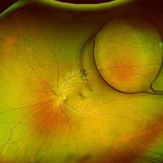
48 year old female referred for pigmented lesion. Exam and photos consistent with well circumscribed CHRPE with lacunae. Patient previously unaware. Observation recommended.
Photographer: Virginia Gebhart, Retina Consultants of Carolina
Imaging device: Optos California
Condition/keywords: CHRPE, congenital hypertrophy of the retinal pigment epithelium (CHRPE)
-
 Best Disease
Dec 9 2025 by Kimberly Wakester
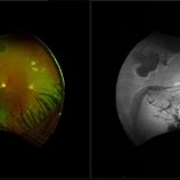
Optomap RBG and AF photograph of an 65-year-old man with Best disease in the left eye. The hypopigmented lesions appear stable on clinical exam and fundus photos compared to previous images. Patient is to continue yearly follow up care with dilated exam and repeat imaging.
Photographer: Kimberly Wakester, COA, OCT-C
Imaging device: Optos California
Condition/keywords: Best Disease, Dystrophies of the Retinal Pigment Epithelium
-
 Dislocated Lens
Dec 8 2025 by Parnian Arjmand, MD, MSc, FRCSC, DABO
A high myope patient presented 12 years after PPV a Cataract extraction for a retinal detachment repair with a new onset of vision loss. A dislocated IOL was noted on clinical examination.
Condition/keywords: Aphakia, dropped IOL, myopia, zonular dehiscence
-
 IOL Drop
Dec 4 2025 by surabhi gupta
A 60 year old man presented with sudden dimunition of vision in right eye. His visual acuity was finger counting at 1 meter and best corrected visual acuity with +10 D was 6/9. Patient was diagnosed with spontaneous right eye IOL bag complex drop in vitreous cavity with superior HST and inferotemporal hole secondary to posterior vitreous detachment . Right eye montage color fundus photo shows rigid IOL bag complex in vitreous cavity with barraged superior HST and inferotemporal hole. Post barrage laser patient underwent pars plana vitrectomy with IOL explantation and scleral fixated IOL.
Photographer: Dr Surabhi Gupta
Imaging device: EDION FA
Condition/keywords: IOL drop
-
 Gyrate Atrophy
Nov 22 2025 by Gaurav Kamble
A 12-year-old female presented with progressive blurring of vision for distance and had a known history of convulsions. Ocular examination revealed bilateral proptosis and megalocornea. Fundus evaluation showed well-defined scalloped areas of peripheral chorioretinal degeneration characteristic of gyrate atrophy, along with cystoid macular edema involving the macular region. The overall clinical picture was consistent with gyrate atrophy.
Photographer: Ms. Vishaka Shah , Isha Eye Care Pvt Ltd ,Khadakpada, Kalyan
Imaging device: Optos Imaging Daytona
Condition/keywords: gyrate atrophy
-
 Falciform Retinal Detachment
Nov 22 2025 by rohan jain
Granular fundus with sclerosed retinal vessels with falciform retinal detachment.
Photographer: Dr. ROHAN JAIN
Imaging device: mirante
Condition/keywords: familial exudative vitreoretinopathy (FEVR), persistent fetal vasculature (PFV), persistent hyperplastic primary vitreous (PHPV), Persistent Hyperplastic Primary Vitreous Fibrovascular membrane, ROP
-
 Starstruck by Stargardt
Nov 17 2025 by SHRADDHA RAJ SHRIVASTAVA
Left eye G-FAF image of a 26 year old patient diagnosed with Stargardt Disease, showing hyperautofluorescent flecks of increased lipofuscin accumulation and dark areas of hypoautofluorescence representing retinal pigment epithelium (RPE) atrophy.
Photographer: Dr. Shraddha Raj Shrivastava
Imaging device: Nidek Mirante SLO/OCT (Confocal scanning/Spectral domain OCT)
Condition/keywords: fleck dystrophy, fundus autofluorescence (FAF), hereditary macular dystrophy, heredomacular degeneration, lipofuscin, Stargardt Disease
-
 Twin Maps of Ischemia: A Fluorescein–OCT Angiography Mirror in Post-Transplant Vasculopathy
Nov 13 2025 by Guilherme Sturzeneker, MD, MSc
Fluorescein angiography (top) and optical coherence tomography angiography (bottom) from both eyes of a 29-year-old woman, obtained 49 days after haploidentical hematopoietic stem cell transplantation, demonstrating marked bilateral macular hypoperfusion with perivascular leakage. OCT-A confirms extensive capillary dropout in both eyes. These findings are consistent with atypical bilateral ischemic retinal vasculopathy unrelated to graft-versus-host disease. Management included oral corticosteroids and intravitreal anti-VEGF injections, with partial anatomical and functional response.
Photographer: Patrick Oikawa, IPEPO - Instituto da Visão
Imaging device: Intalight Dream OCT
Condition/keywords: Fluorescein angiography, Hematopoietic stem cell transplantation, Ischemic retinal vasculopathy, Non-graft-versus-host disease, OCT Angiography, oncology
-
 The Great Disc-guise
Nov 12 2025 by SHRADDHA RAJ SHRIVASTAVA
Right eye pseudocolor fundus photo of a 20 year old with Both eyes Pathological Myopia (spherical refractive error of - 18.00 DS in BE), showing a tilted myopic disc with peripapillary atrophy, and extensive posterior staphyloma baring the underlying choroidal vessels and scleral tissue. We can also see a well-defined round chorioretinal atrophic (CRA) patch superonasal to the disc, giving the illusion of double disc on cursory fundus examination.
Photographer: Dr. Shraddha Raj Shrivastava
Imaging device: Nidek Mirante SLO/OCT (Confocal scanning/Spectral domain OCT)
Condition/keywords: chorioretinal atrophy, High Myopia, pathologic myopia, peripapillary atrophy, posterior staphyloma
-
 Unilateral Pigmentary Retinopathy
Nov 9 2025 by Hrishikesh Naik, MS
Montage fundus photographs of a 47 year old female presenting with unilateral vision loss in the left eye. Fundoscopy revealed extensive intraretinal pigment clumps, waxy disc pallor, and marked vessel attenuation in the left eye with a normal fundus in the right. Electroretinography showed unilateral reduction in rod and cone function. Unilateral pigmentary retinopathy, an uncommon variant of retinitis pigmentosa (reported incidence ˜ 5%) presents with RP-like changes in one eye, the fellow eye being completely normal. Proposed causes include lyonization and somatic mosaicism. Conditions which mimic RP should be excluded, and any diagnoses should be supported with electrodiagnostic tests and autofluorescence imaging. Management parallels RP, focusing on cataract and macular complications and long-term follow-up to monitor possible bilateral progression.
Imaging device: Zeiss Visucam 224
Condition/keywords: montage, retinitis pigmentosa, unilateral
-
 Retinitis Pigmentosa: Now available in its Pericentral edition
Nov 7 2025 by SHRADDHA RAJ SHRIVASTAVA
Left eye Green-FAF image, of a 50 year old patient, diagnosed with bilateral Pericentral variant of Retinitis Pigmentosa. The disease is characterized by pigmentary changes closer to the macula, and an earlier involvement of central visual acuity as compared to typical RP. We can see prominent, scalloped hypoautofluorescent lesions in the pericentral region, which corresponds to areas of severe RPE atrophy and photoreceptor cell loss. Macula shows preserved background autofluorescence, with darker areas corresponding to no detectable fluorescence due to macular atrophy (loss of melanin/lipofuscin).
Photographer: Dr. Shraddha Raj Shrivastava
Imaging device: Nidek Mirante SLO/OCT (Confocal scanning/Spectral domain OCT)
Condition/keywords: ATYPICAL RETINITIS PIGMENTOSA, fundus autofluorescence (FAF), pericentral retinitis pigmentosa, RP variant
-
 Retinitis Pigmentosa: Now available in its Pericentral edition
Nov 7 2025 by SHRADDHA RAJ SHRIVASTAVA
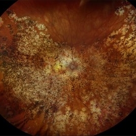
Right eye fundus photo of a 50 year old patient, diagnosed with bilateral Pericentral variant of Retinitis Pigmentosa. True to the subtype, the pigmentation is closer to fixation. There are bony spicules like pigmentary changes and RPE atrophy seen around the macula and disc (posterior pole), just adjacent to the arcades, while the peripheral fundus appears unaffected. The macula shows severe macular atrophy and scarring. Similar changes were observed in the left eye.
Photographer: Dr. Shraddha Raj Shrivastava
Imaging device: Nidek Mirante SLO/OCT (Confocal scanning/Spectral domain OCT)
Condition/keywords: pericentral retinitis pigmentosa, retinitis pigmentosa (RP) dystrophy, Rod cone dystrophy, RP variant
-
 Gravity = 1, Zonules = 0 : Cionni ring-IOL-Bag complex subluxation
Nov 7 2025 by SHRADDHA RAJ SHRIVASTAVA
Right eye anterior segment slit-lamp image of a 30 year old male, who was operated for spontaneous bilateral inferior subluxation of crystalline lens. Primary surgery was performed almost 20 years ago in which lens extraction was done followed by IOL placement in bag after stabilising it with Cionni ring. The patient presented to us recently with right eye diminution of vision and was noted to have inferiorly subluxated IOL-capsular bag complex, with vitreous in AC coming from the superior aphakic area. Interestingly, we have also captured in this image - the capsular tension ring (Cionni ring) with its central fixation eyelet.
Photographer: Dr. Shraddha Raj Shrivastava
Condition/keywords: dislocated IOL, dropped capsular IOL bag complex, IOL drop, Subluxated IOL, zonular dehiscence
-
 All That Glows Yellow Isn’t Mellow: Coats' Disease Unveiled
Nov 4 2025 by SHRADDHA RAJ SHRIVASTAVA
Montage fundus image of an 11 year old boy diagnosed with left eye Coats' disease (stage 3A1), reveals a hyperemic disc and surrounding intra-retinal exudates superior to the disc. There is a single fibroglial nodule at the macula causing submacular fibrosis with exudation. We can see areas of pigmentary changes and RPE atrophy in posterior pole and mid-peripheral retina supero-temporally. There is massive yellowish subretinal exudation in all the quadrants, which are associated with telangiectatic aneurysmal capillary dilation, more prominently seen in the nasal periphery. Supero-nasally we can also see an orange-red elevated vaso-proliferative mass with overlying dilated capillaries, which has likely developed secondary to untreated long standing disease. We can also see associated extrafoveal subtotal exudative retinal detachment in the inferior and nasal quadrants.
Photographer: Dr. Shraddha Raj Shrivastava
Imaging device: Nidek Mirante SLO/OCT (Confocal scanning/Spectral domain OCT)
Condition/keywords: COATS DISEASE, exudative detachment, leukocoria, subretinal exudates, Xanthocoria, yellow exudate
-
 Optic Disc Drusen in Rod Cone Dystrophy
Nov 3 2025 by Malvika Singh
Fundus autofluorescence of a 22 year old male with rod cone dystrophy with hyperautofluorescent disc drusen.
Photographer: Dr Malvika Singh, Retina Foundation, Ahmedabad, India
Imaging device: Mirante SLO/OCT
Condition/keywords: optic disc drusen, Rod cone dystrophy
-
 Pigmentary Retinal Dystrophy
Oct 30 2025 by Kimberly Wakester
Optomap RGB of an 77-year-old-woman with Pigmentary Retinal Dystrophy in the left eye. Patient is to continue follow up care yearly with dilated exam and diagnostic testing.
Photographer: Kimberly Wakester, COA, OCT-C
Imaging device: Optos California
Condition/keywords: bone spicules, Pigmentary Retinal Dystrophy
-
 ERMageddon - Wrinkle in the Space-time Fabric of Macula
Oct 29 2025 by SHRADDHA RAJ SHRIVASTAVA
38 year old female with Epiretinal Membrane (ERM) over macula, post laser barrage for multiple symptomatic Horse-shoe Tears (HSTs) and Lattice Degenerations (seen on wide-field image). Posterior pole revealed tilted disc with peripapillary atrophy. There is thick opaque epiretinal membrane obscuring the underlying superior arcade vessels and causing foveal ectopia with distortion of perimacular vasculature. Patient was planned for Right Eye pars plana vitrectomy for ERM peeling.
Photographer: Dr. Shraddha Raj Shrivastava
Imaging device: Nidek Mirante SLO/OCT (Confocal scanning/Spectral domain OCT
Condition/keywords: BARRAGE LASER, ectopic fovea, epiretinal membrane (ERM), horseshoe tear, lattice degeneration, vitreomacular traction (VMT)
-
 ERMageddon - Wrinkle in the Space-time Fabric of Macula
Oct 29 2025 by SHRADDHA RAJ SHRIVASTAVA
38 year old female with Epiretinal Membrane (ERM) over macula, post laser barrage for multiple symptomatic Horse-shoe Tears (HSTs) and Lattice Degenerations. Posterior pole revealed tilted disc with peripapillary atrophy. There is thick opaque epiretinal membrane obscuring the underlying superior arcade vessels and causing foveal ectopia with distortion of perimacular vasculature. Patient was planned for Right Eye pars plana vitrectomy for ERM peeling.
Photographer: Dr. Shraddha Raj Shrivastava
Imaging device: Nidek Mirante SLO/OCT (Confocal scanning/Spectral domain OCT
Condition/keywords: ectopic fovea, epiretinal membrane (ERM), ERM, horseshoe tear, vitreomacular traction (VMT)
-
 Retinopathy of Prematurity
Oct 27 2025 by Anjana Mirajkar, MS Ophthalmology
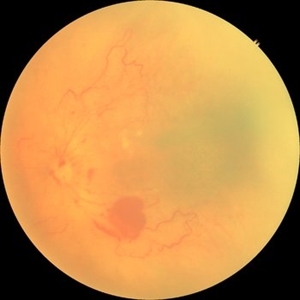
Fundus photograph of a premature baby showing flat neovascularization with looping of the vessels with bleed in zone 1/2 with plus disease suggestive of A-ROP.
Photographer: Dr. Anjana Mirajkar- HV desai eye hospital ,Pune
Imaging device: Retcam
Condition/keywords: aggressive posterior retinopathy of prematurity (APROP)
-
 Retinopathy of Prematurity
Oct 26 2025 by Anjana Mirajkar, MS Ophthalmology
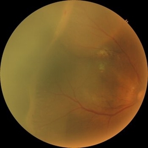
Fundus photograph of right eye of premature baby showing stage 3 in zone 2 posterior.
Photographer: Dr. Anjana Mirajkar- HV desai eye hospital ,Pune
Imaging device: Retcam
Condition/keywords: retinopathy of prematurity (ROP), stage 3
-
 Retinopathy of Prematurity
Oct 26 2025 by Anjana Mirajkar, MS Ophthalmology
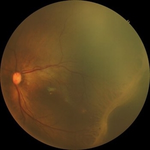
Fundus photograph of a left eye of a premature baby showing stage 3 in zone 2 posterior.
Photographer: Dr. Anjana Mirajkar- HV desai eye hospital ,Pune
Imaging device: Retcam
Condition/keywords: retinopathy of prematurity (ROP), retinopathy of prematurity stage 3
-
 Retinopathy of Prematurity
Oct 26 2025 by Anjana Mirajkar, MS Ophthalmology
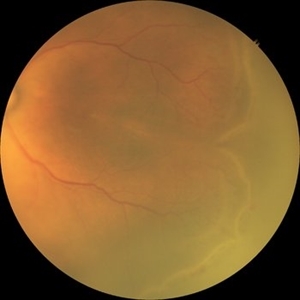
Fundus photograph of left eye premature baby having stage 3 in zone 2A with a secondary notch.
Photographer: Dr. Anjana Mirajkar- HV Desai eye hospital ,Pune
Imaging device: retcam
Condition/keywords: retinopathy of prematurity (ROP), stage 3
-
 Posterior Dislocated Intraocular Lens
Oct 23 2025 by Aditya S Kelkar, MS, FRCS, FASRS,FRCOphth
Fundus photograph of an 53-year-old man with a posteriorly dislocated intraocular lens near the posterior pole.
Photographer: Dr Tejal Rao, National Institute of Ophthalmology, Pune, India
Imaging device: Optos Daytona
Condition/keywords: dislocated intraocular lens (IOL), IOL drop
-
 Radiation Retinopathy with Rhegmatogenous Retinal Detachment
Oct 20 2025 by Meng-Hsin Chen
Fundus photo of a 54-year-old woman showing chronic radiation retinopathy from in-utero exposure with rhegmatogenous retinal detachment at the 3:00 -9:00 region, atrophic retina, and PVR. Lattice degeneration is present superiorly and inferiorly with a neovascular frond.
Photographer: Meng-Hsin Chen
Condition/keywords: atrophic retina, lattice degeneration, neovascular frond, proliferative vitreoretinopathy (PVR), radiation retinopathy, retinal detachment
-
 Multifocal Choroiditis with Panuveitis
Oct 16 2025 by Virginia Gebhart
39 year old female diagnosed with MCP in 2009. Extensive RPE changes and hypertrophy, arterial attenuation and pale nerve. Currently no active inflammation.
Photographer: Virginia Gebhart, Retina Consultants of Carolina
Imaging device: Optos California
Condition/keywords: corticosteroid-induced glaucoma, hypertrophy, multifocal chorioretinitis (MCP), PALE DISC
 Loading…