Search results (59 results)
-
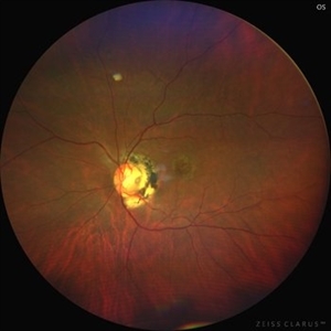
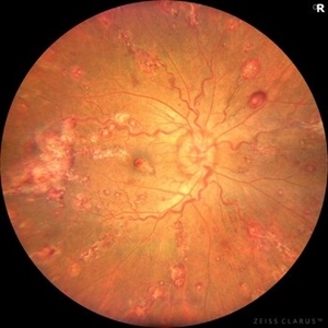 Leukemic Retinopathy and Optic Neuropathy
Aug 25 2025 by Elysse Tom, MD
Fundus photo of a 45-year-old woman with chronic myeloid leukemia.
Condition/keywords: leukemia, leukemic infiltration, Leukemic optic neuropathy
-
 Presumed Congenital Toxoplasmosis
Aug 16 2025 by Vishal Agrawal, MD, FRCS,FACS,FASRS
Fundus picture of 7 a year-old boy with esotropia. OCT showed complete atrophy & disorganization of the overlying RPE and neurosensory retina.
Photographer: Dr Ayushi Gupta
Imaging device: Clarus 700
Condition/keywords: coloboma of macula, toxoplasmosis
-
 CHRPE and Bear Tracks
Jan 7 2025 by Drew Mitchell
Fundus Autofluorescence of a CHRPE and Bear Tracks.
Photographer: Drew Mitchel, OCT-C
Imaging device: Optos Silverstone
Condition/keywords: bear tracks, CHRPE, congenital hypertrophy of the retinal pigment epithelium (CHRPE)
-
 MIDD (Maternally Inherited Diabetes and Deafness) - Right AF
Nov 30 2024 by John S. King, MD
Both right and left eyes have symmetrical ring of mottled hypo/hyper AF around the fovea and disc. The HyperAF areas correspond to RPE deposits on OCT as well as areas of blockage on FA, and drusenoid deposits seen on fundus photos. Disc drusen in right eye present as HyperAF spot 57 yo WF referred for AMD vs Pattern Dystrophy that was diagnosed 10 years ago. Reported some slow progressive vision loss in both eyes for distance and near. Denies nyctalopia or hemeralopia. Background medical history includes HTN, CVD, and DM. No family history of eye problems. Denied pentosan use. Anterior segment showed moderate cataracts (OD>OS). Posterior segment exam showed macular changes and mild NPDR. The macular appearance showed a symmetrical, paramacular ring of fleck-like drusenoid material with some faint focal areas of RPE hyperplasia. Fundus Photos, AF, OCT were performed as well as a gene test. Further questioning showed revealed that her mother and maternal grandmother had both diabetes mellitus and sensorineural hearing loss. The patient developed diabetes in her teens, and some high frequency hearing loss in her early twenties. She had not had a previous genetic test or diagnosis of MIDD. Gene testing is pending for the mitochondrial component. Invitae's retinal panel, which does not include mitochondrial disorders, only showed a variant of uncertain significance, HMCN1. I discussed this case with Dr. Freund, and it is similar to a the case report : Inoue M, Kiss S, Freund KB. MACULAR PIGMENT RINGS AS THE PRESENTING FINDING OF MITOCHONDRIAL MYOPATHY, ENCEPHALOPATHY, LACTIC ACIDOSIS, AND STROKELIKE EPISODES. Retin Cases Brief Rep. 2015 Fall;9(4):260-4. doi: 10.1097/ICB.0000000000000182. PMID: 26200388.
Photographer: Grace Melton and Carley Gunn
Imaging device: Clarus
Condition/keywords: Macular Dystrophy, Maternally Inherited Diabetes and Deafness, MIDD, Mitochondrial Disorder
-
 Pericentral Retinitis Pigmentosa
Sep 6 2024 by Mauricio Bayram-Suverza, MD
A 65-year-old male patient reports experiencing bilateral blind spots that have gradually intensified over time. Genetic testing was unrevealing. The fundus autofluorescence image shows a hypoautofluorescent ring in the posterior pole, especially nasal to the nerve and along arcades.
Photographer: Mauricio Bayram-Suverza, Casey Eye Institute, OHSU.
Imaging device: Optos California
Condition/keywords: fundus autofluorescence (FAF), inherited retinal disease, nyctalopia, retinal dystrophy, retinitis pigmentosa
-
 B-FAF in Stargardt's Disease
Jul 4 2024 by Tejaswita Verma
Blue fundus autofluorescence showing hypoautofluorescence picture of a 28 year old male with 6/60 vision in BE in a case of Stargardt's disease.
Photographer: DR. TEJASWITA VERMA
Imaging device: MIRANTE
Condition/keywords: fundus autofluorescence (FAF), hereditary macular dystrophy, Stargardt disease
-
 Dislocated Crystalline Lens
Mar 19 2024 by Annaka Gooding
Ultra Wide field fundus photography of a 70 year old male who presented to clinic with a sudden increase of vision due to dropped crystalline lens secondary to severely dense cataract. Patient reported seeing a full black circle in his inferior visual field. Patient's visual acuity at time of visit was 20/100 with a +5.00 diopter lens. The physician recommended surgical intervention, and discussed surgery for PPV/PPL/IOL implantation with an ACIOL.
Photographer: Annaka Gooding, CPO
Imaging device: Optos California RGB
Condition/keywords: dislocated crystalline lens, fundus photography, inferior retina, OPTOS CALIFORNIA RGB, Right Eye, Ultra-wide field retinal imaging
-
 Venous Loop
Feb 20 2024 by Soobien Lee
A 77-year-old male with a history of bilateral optic neuropathy from bilateral optic nerve sheath meningiomas S/P radiation/proton-beam therapies. Presented with radiation retinopathy OS and a known venous loop OS.
Photographer: Gavin Bragdon, Elman Retina Group
Imaging device: Optos Ultra-Widefield Fluorescein Angiography
Condition/keywords: fluorescein angiogram (FA), Optos, radiation retinopathy, retinal vascular disease, venous loop
-
 New Retinal Detachment 6w s/p RD repair
Nov 16 2023 by Virginia Gebhart
13 year old male presented with new blind spot 6 weeks s/p RD repair with cryo/scleral buckle/prophylaxis laser with gas bubble. New RD involving the macula, posterior to scleral buckle, secondary to PVD. Small gas bubble remaining. Pt was brought back to OR for repeat PPV and silicone oil repair
Photographer: Virginia Gebhart
Imaging device: Optos
Condition/keywords: gas bubble, Retinal Detachment, retinal detachment of the macula, scleral buckle
-
 Wyburn -Mason Syndrome
Jul 21 2023 by Rajshree Arora
9 year old girl presented with unilateral pulsatile mild to moderate proptosis with lost vision.
Photographer: Dr Rajshree Arora,Dr Neelam Choksi
Condition/keywords: Wyburn -Mason Syndrome
-
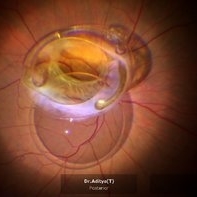 Rescuing IOL CTR Bag Complex
Jun 14 2023 by Aditya S Kelkar, MS, FRCS, FASRS,FRCOphth
INTRAOPERATIVE SNAPSHOPT IN ZEISS ARTEVO 800 OF DROPPED IOL CTR BAG COMPLEX IN A 71 YEAR OLD MALE PATIENT
Photographer: SUBHASREE DUTTA, NATIONAL INSTITUTE OF OPHTHALMOLOGY, PUNE
Imaging device: ZEISS ARTEVO 800
Condition/keywords: dropped capsular IOL bag complex
-
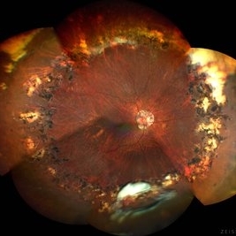
 Gyrate Atrophy
Apr 12 2023 by Ahmed Abbas Hashmi, OD
Left eye fundus of a 53-year-old male patient with advanced gyrate atrophy of the choroid and retina with macular sparing. Optic nerve head is healthy.
Photographer: Ahmed Abbas Hashmi
Imaging device: Topcon TRC-NW8F
Condition/keywords: chorioretinal atrophy
-
 Solitary large Congenital Hypertrophy of Retinal Pigment Epithelium (CHRPE)
Jul 1 2023 by Aditya S Kelkar, MS, FRCS, FASRS,FRCOphth
Right eye fundus photograph of a 42 year old asymptomatic male demonstrating a superotemporal solitary large Congenital Hypertrophy of Retinal Pigment Epithelium (CHRPE) lesion.
Photographer: Optom Komal Jangam
Imaging device: OPTOS DAYTONA
Condition/keywords: CHRPE
-
 PEHCR (Peripheral Exudative Hemorrhagic Chorioretinopathy)
May 12 2023 by Niloofar Piri, MD
Ultrawide fundus photograph of the left eye demonstrating extensive peripheral hemorrhagic exudative detachment in a 79 yo Caucasian female with prior history of non-exudative AMD. Recent diagnosis of Acute myeloid leukemia with low platelet count which might have contributed to the above presentatuon. Please note the temporal subretinal hemorrhage as well as RPE atrophy and hyperplasia in the macula.
Photographer: Rocio Bentivegna, MD, Saint Louis University; Jessica Maddox, COA, Saint Louis University
Condition/keywords: peripheral exudative hemorrhagic chorioretinopathy (PEHCR)
-
 North Carolina Macular Dystrophy collage
Nov 21 2022 by Kent W. Small, MD, FASRS, FACS
Color fundus photo collage of 24 members of a single family with NCMD chr6: 99593030G>T
Condition/keywords: North Carolina Macular Dystrophy (MCDR1)
-
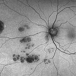
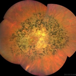
 Rod Cone dystrophy
Nov 29 2022 by Niloofar Piri, MD
Fundus autofluorescence of the left eye in a 58 yo male with rod cone dystrophy. He presented with night blindness and peripheral vision loss since youth and recent decrease in central vision for the past 10 years. Notice multiple coin shaped hypoautofluorescent pacthes within central 20 degrees which are coalescing centrally. (fundus photo uploaded separately) He has one pathogenic variants of both CEP290 and PRPH2 genes.
Photographer: Sean Kelso, Saint Louis University
Condition/keywords: hereditary retinal degeneration, hereditary retinal dystrophy, rod cone dystrophy
-
 Angioid Streaks
Dec 14 2022 by Pramod Kumar Suman, MBBS, MD
Fundus autofluorescence photograph of a 65-year-old male with numerous narrow, irregular streaks radiating in a circumferential pattern within the posterior pole with macular atrophy.
Photographer: Pramod Kumar Suman, Retina Foundation, Ahmedabad
Imaging device: Mirante
Condition/keywords: angioid streaks
-
 Posteriorly dislocated IOL
Oct 22 2022 by Vishal Agrawal, MD, FRCS,FACS,FASRS
67 yr old male , post PPV for retinal detachment ( 5 years ) presented with sudden DOV . On examination posteriorly dislocated 4 loop haptic iol - bag complex was noted .
Photographer: Pankaj
Imaging device: CLARUS 700
Condition/keywords: dropped intraocular lens (IOL)
-
 Tapetoretinal Degeneration
Sep 7 2022 by JEFFERSON R SOUSA, Tecg.º (Biomedical Systems Technology)
Patient 52 years old, Male, progressive loss of vision since the age of 20. Retinography showed mobilization of pigments in osteoblasts, extensive area of atrophy of the pigmentary epithelium and choroid. On fluorescein angiography, typical changes following the characteristic patterns of paracentra retinal retinitis pigmentosa. Autofluorescent fundus with a sectorial autohypofluorescence pattern in the regions of atrophies.
Photographer: JEFFERSON ROCHA DE SOUSA - Retinal Department at Instituto Dr. Suel Abujamra Sao Paulo-Brazil
Imaging device: Clarus 700 - Zeiss, composite of four 135 degree images.
Condition/keywords: pericentral retinitis pigmentosa, tapeoretinal degeneration
-
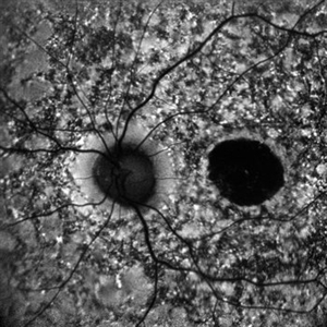
 Rod cone dystrophy autofluorescence
Sep 19 2022 by Kenneth Fong
34 year old male with colour blindness and loss of visual field
Condition/keywords: retinal dystrophy
-
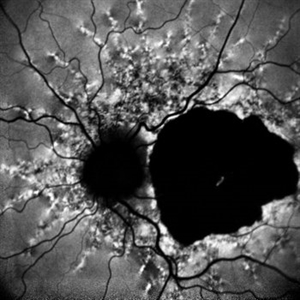
 Leukemic optic neuropathy
Oct 28 2022 by pedro fernandes souza neto
Fundus photograph of an 18-year-old woman with Leukemic optic neuropathy.
Photographer: Pedro Fernandes, Universidade Federal da Bahia
Condition/keywords: Leukemic optic neuropathy
-
 CERKL-related Cone Rod Dystrophy
Jun 27 2022 by Hanna Choi
37-year-old female with cone-rod dystrophy. Developed photophobia and progressive blurry vision in the third decade. VA 20/40 OD, 20/30 OS. The patient is compound heterozygous for pathogenic mutations in the CERKL gene (Arg465Trp and Arg283*).
Photographer: Kaitlynn Silva, New England Retina Consultants
Imaging device: Ultrawide-field Optos Fundus Photography, Autofluorescence, Fluorescein Angiography
Condition/keywords: cone dystrophy, inherited retinal disease, maculopathy
-
 Spontaneously Dropped Lens in a Congenital Rubella Syndrome
Apr 30 2022 by NEIFFER RABELO
Intraoperative photograph of a 68-year-old patient with congenital rubella syndrome and light perception visual acuity since childhood. The image shows a pigmentary retinopathy and the lens spontaneously displaced into the vitreous cavity. The patient sought care complaining of a total and sporadic loss of vision that was hindering her in daily tasks. Surgery was indicated to remove the lens.
Photographer: Rodrigo Dos Anjos Versiani - Retina Institute - Belo Horizonte - Brazil
Imaging device: ZEISS OPMI LUMERA 700
Condition/keywords: dropped nucleus, retina surgery, rubella retinopathy
-
 Thioridazine-toxicity
Apr 30 2022 by Niloofar Piri, MD
61 yo male with PMH of longstanding schizophrenia since 20s with secondary intellectual disability presented with decreased vision following a recent stroke. He was found to have bilateral chorio-retinal atrophy involving posterior pole with scalloped edges and coin shaped atrophic area at margins extending into mid-periphery, diagnosis most concerning for intermediate stage thioridazine toxicity given the history. Mother could find handwritten prescriptions from 1990s when he was on Thioridazine 800 mg daily for unknown period of time. Patient had better vision in the left eye which was affected by recent stroke and prompted him to seek medical care. Fundus photograph of the right eye is demonstrated here.
Photographer: Jacob Grodsky, MD
Condition/keywords: drug toxicity, thioridazine toxicity, toxic retinopathy
-
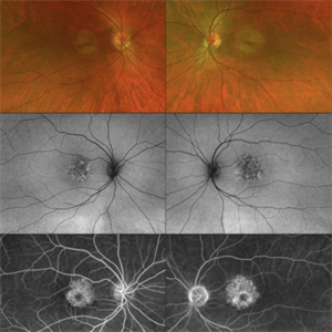
 Optic disc pit
Mar 21 2022 by T. P . VIGNESH, MBBS,MS
Fundus photo of Left eye of a 55 year male patient revealing optic disc pit with temporal barrage laser marks and foveal schisis with RPE atrophic changes.
Photographer: Bharathi Singaravel
Imaging device: Zeiss Clarus
Condition/keywords: Optic disc pit
 Loading…