Search results (2916 results)
-
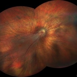
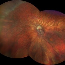
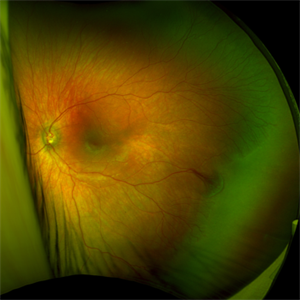
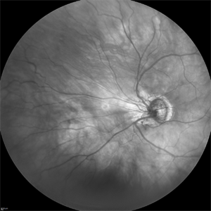
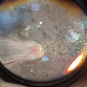
 Falciform Retinal Detachment
Nov 22 2025 by rohan jain
Granular fundus with sclerosed retinal vessels with falciform retinal detachment.
Photographer: Dr. ROHAN JAIN
Imaging device: mirante
Condition/keywords: familial exudative vitreoretinopathy (FEVR), persistent fetal vasculature (PFV), persistent hyperplastic primary vitreous (PHPV), Persistent Hyperplastic Primary Vitreous Fibrovascular membrane, ROP
-
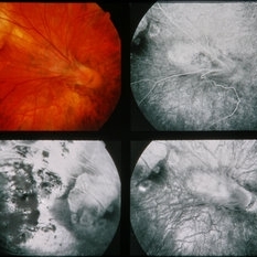
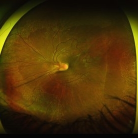
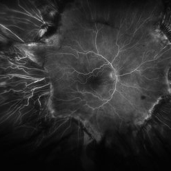 Fluorescein Angiogram of ROP With Cryo Scarring
Jul 7 2025 by Jenn Geelan
FA photo of a 34 year old male with prior stage 3 ROP with history of 360 degree cryotherapy.
Photographer: Jenn Geelan, Retina-Vitreous Surgeons of CNY
Imaging device: Optos California
Condition/keywords: cryotheraphy scar, fluorescein angiogram (FA), fundus photograph, retinopathy of prematurity (ROP), ROP, tilted disc
-
 Macular Dragging
Dec 12 2024 by César Adrián Gómez Valdivia, MD
Macular dragging found in a 14 year-old female patient with ROP history. Findings were bilateral.
Photographer: @eyemissu2
Imaging device: TOPCON TRC-50DX
Condition/keywords: macular dragging, rop, ROP MACULAR DRAGGING
-
 Macular dragging OD due to cicatricial ROP
Mar 2 2023 by Kamal Kishore, MD, MBBS
A 21-year-old female with macular dragging OD secondary to untreated cicatricial ROP.
Photographer: Jessi Wright
Imaging device: Zeiss Clarus
Condition/keywords: macular dragging, rop
-
 Macular dragging OS in cicatricial ROP
Mar 2 2023 by Kamal Kishore, MD, MBBS
21-year-old with macular dragging due to untreated cicatricial ROP. Note large peripheral avascular retina (PAR) at the nasal periphery.
Photographer: Jessi Wright
Imaging device: Zeiss Clarus
Condition/keywords: macular dragging, rop
-
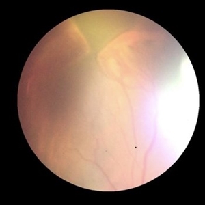 ROP
ROP
-
 ROP
ROP
Sep 4 2018 by PAVEL FLORES-MORENO
ROP-Stage 2. Zone 2.
Photographer: Pavel Flores
Condition/keywords: retinopathy of prematurity (ROP)
-
 ROP
ROP
Sep 4 2018 by PAVEL FLORES-MORENO
ROP-Stage 2. Zone 2.
Photographer: Pavel Flores
Condition/keywords: retinopathy of prematurity (ROP)
-
 ROP
ROP
May 15 2025 by Max D Schlesinger, MD
Fundus Photo of an ex-25 week premature infant, now 52 weeks gestational age, with persistent avascular retina and popcorn hemorrhage.
Photographer: Paul Whitten
Condition/keywords: ROP
-
 ROP
ROP
Feb 13 2014 by Howard Schatz, MD
39-year-old white male. RE 10/200 LE 20/30. Retinopathy of prematurity.
Condition/keywords: retinopathy of prematurity (ROP)
-
 ROP
ROP
Feb 13 2014 by Howard Schatz, MD
35-year-old female. RE 20/100 LP. Retinopathy of prematurity.
Condition/keywords: retinopathy of prematurity (ROP)
-
 ROP
ROP
Feb 13 2014 by Howard Schatz, MD
37-year-old white female. RE 20/40 LE CF1'. Retinopathy of prematurity.
Condition/keywords: retinopathy of prematurity (ROP)
-
 ROP
ROP
-
 ROP
ROP
Mar 26 2025 by Korey Starkey
9 month old patient presents today with Retinopathy of Prematurity in both eyes. Patient was born at gestational age of 25 weeks 2 days, 940g. Left eye presents with vitreoretinal traction and peripheral VH with regressed stage 3 and persistent stage 2 disease.
Photographer: Korey Starkey
Imaging device: Optos
Condition/keywords: retinopathy of prematurity stage 2, rop, stage 3, vitreoretinal traction, vitreous hemorrhage
-
 ROP stage 3
Sep 22 2022 by Filip Kecer
IR cSLO widefield image of an 27-year-old man born as premature
Photographer: Filip Kecer, National Institute of Childrens Diseases
Imaging device: Spectralis, Heidelberg Engineering
Condition/keywords: retinopathy of prematurity stage 3, rop
-
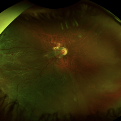
 ROP / Disk Dragging
May 27 2025 by César Adrián Gómez Valdivia, MD
Macular dragging found in a 14 year-old female patient with ROP history. Findings were bilateral.
Photographer: @eyemissu2
Imaging device: California ICG OPTOS
Condition/keywords: Disk Dragging
-
 ROP 4B late Retinal Findings
Mar 31 2022 by Franco Benvenuto, MD
A 9-year-old male, that was born at 30 weeks of gestation with birth weight of 1500 g and history of hospitalization for 20 days with respiratory distress and packed red blood cell transfusion for anemia. At the first exam, both eyes were with stage 4B ROP. Vitrectomy with 25 G was done in both eyes. The flat fibrosis dragged the macula nasally in both the eyes.
Photographer: Franco Benvenuto, Universidad de Buenos Aires, Argentina; Universidad de Guadalajara, México.
Condition/keywords: cicatricial retinopathy of prematurity, retinopathy of prematurity (ROP)
-
 ROP 5A
Jan 24 2022 by Alexandre Grandinetti, MD, PhD
ROP retinal detachment.
Photographer: Alexandre Grandinetti
Imaging device: RetCam
Condition/keywords: retinopathy of prematurity (ROP), total retinal detachment
-
 ROP Dragging
-
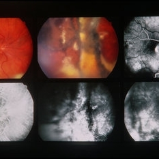
 ROP FA OD
Apr 27 2018 by Brenda Fallas
4-month-old baby with regressed ROP post-Avastin.
Photographer: Brenda Fallas, Bascom Palmer Eye Institute, Miami, FL
Imaging device: RETCAM III 130 degree lens mongtage
Condition/keywords: FA late phase leakage, fluorescein angiogram (FA), retina, retinopathy of prematurity (ROP)
-
 ROP FA OS
Apr 27 2018 by Brenda Fallas
4-month-old baby with regressed ROP post-Avastin.
Photographer: Brenda Fallas, Bascom Palmer Eye Institute, Miami, FL
Imaging device: RETCAM III 130 degree lens montage
Condition/keywords: FA late phase leakage, fluorescein angiogram (FA), retinopathy of prematurity (ROP)
-
 ROP Leukocoria
Oct 19 2012 by Larry Halperin, MD
ROP leukocoria
Condition/keywords: leukocoria, retinopathy of prematurity (ROP)
-
 ROP related retinal detachment
Jun 8 2022 by Alexandre Grandinetti, MD, PhD
16-year-old girl with macula off retinal detachment and ROP cicatricial changes. She was born with 28 weeks of gestation and had no treatment
Photographer: Corina Skrzek, Hospital de Olhos do Paraná
Imaging device: California
Condition/keywords: rop retinal detachment
-
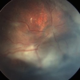
 ROP SEQUELAE
Apr 26 2023 by Kalyan Singh
Fundus photograph of an young boy with history of premature birth.
Photographer: Dr Kalyan Singh, Junior resident , Department of ophthalmology, GSVM MEDICAL COLLEGE KANPUR
Imaging device: One plus 10 R ( smartphone)
Condition/keywords: ROP MACULAR DRAGGING
-
 ROP-Zone-I-Stage-3-Plus
Jun 3 2022 by Dipak Nag, MBBS, FCPS, MSc, FRF
Fundus photograph of a child of gestational age 26 weeks and birth weight 1050 grams, shows dilatation and tortuosity of vessels in zone I, extra-retinal fibro-vascular proliferation, hemorrhage with huge peripheral avascular area.
Photographer: Dipak Nag, National Institute of Ophthalmology, Dhaka, Bangladesh
Imaging device: RetCam shuttle
Condition/keywords: retinopathy of prematurity (ROP), retinopathy of prematurity Plus disease, retinopathy of prematurity stage 3, retinopathy of prematurity zone I
 Loading…