Search results (1738 results)
-
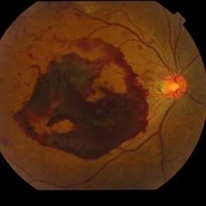
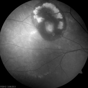
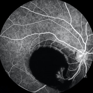 Sub-ILM Hemorrhage with Neovessels
Apr 30 2020 by Saurabh Deshmukh, MBBS, DNB, FVRS, MNAMS
Late arteriovenous phase FA showing a large sub-internal limiting membrane hemorrhage with overlying neovessels. This hypertensive patient presented with a visual acuity of counting fingers at 2 meters. The patient was advised intravitreal anti-VEGF injection, Nd: YAG Membranotomy, and systemic control of hypertension.
Photographer: Saurabh Deshmukh, Sri Sankaradeva Nethralaya, Guwahati, India
Imaging device: Topcon TRC-50 DX
Condition/keywords: hypertensive retinopathy, neovascularization elsewhere (NVE), subILM hemorrhage
-
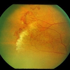
---thumb.jpg/image-square;max$300,300.ImageHandler) Proliferative Diabetic Retinopathy (PDR) & Traction Retinal Detachment
Proliferative Diabetic Retinopathy (PDR) & Traction Retinal Detachment
Feb 13 2013 by From the Collections of Thomas M. Aaberg, MD and Thomas M. Aaberg Jr., MD
Florid NV with early macular TRD.
Condition/keywords: neovascularization (NV), tractional retinal detachment
-
 Sickle Cell Sea Fan Retinopathy
Jun 4 2014 by Henry J. Kaplan, MD
Sea fan peripheral retinal neovascularization in sickle cell anemia.
Condition/keywords: sea fan, sickle cell retinopathy
-
 Venous Loop & Venous Beading
May 31 2014 by Hamid Ahmadieh, MD
Color fundus photograph of the left eye of a diabetic patient with NVD, NVE, venous loop and venous beading.
Photographer: Elham Salehi, Negah Eye Center, Tehran
Condition/keywords: color fundus photograph, neovascularization elsewhere (NVE), neovascularization of the disc (NVD), proliferative diabetic retinopathy (PDR), venous beading, venous loop
-
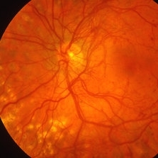
 Proliferative Diabetic Retinopathy
Sep 17 2012 by Michael P. Kelly, FOPS
Retinal fundus photograph of a patient with PDR and NVD.
Photographer: Michael P. Kelly, FOPS Director, Duke Eye Labs, Duke University Hospital, Duke Eye Center
Imaging device: Topcon
Condition/keywords: neovascularization of the disc (NVD)
-
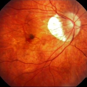
 Proliferative Diabetic Retinopathy with NVD
Oct 4 2012 by Michael P. Kelly, FOPS
Photographer: Michael P. Kelly, FOPS Director, Duke eye Center Labs, Duke University Hospital
Condition/keywords: neovascularization of the disc (NVD), retinal neovascularization
-
 Diabetic Macular Edema, Proliferative Diabetic Retinopathy, Neovascularization Elsewhere, DME, PDR, NVE
Apr 1 2013 by James B. Soque, CRA, OCT-C, COA, FOPS
39-year-old white female and long standing diabetis, c/o new peripheral symptoms of left eye. FA OS reveals diabetic macular edema, microaneurysms, and neovasculaization elsewhere. Fluorescein Angogram, Early Phase, 50 Deg, 2x Mag.
Photographer: James B Soque, CRA, COA
Imaging device: Topcon TRC 50DX with MERGE software, OIS 10.6.45
Condition/keywords: diabetic macular edema, neovascularization (NV), proliferative diabetic retinopathy (PDR)
-
 Myopic CNV
Jan 11 2013 by Alex P. Hunyor, MD
Myopic macular degeneration complicated by subretinal neovascularisation, left eye.
Condition/keywords: high myopia, myopia, myopic choroidal neovascularization (CNV)
-
 Myopic Choroidal Neovascular Membrane
Mar 25 2013 by Ratimir Lazic, MD, PhD
Color fundus photography of a 33-year-old female. In macular area subretinal hemorrhage can be seen. Area of atrophy temporal from PNO. Myopic changes of posterior pole and mid periphery can be noticed. The patient has been treated with 2 consecutive ranibizumab intravitreal injections. BCVA at baseline was 0,05 (Snellen lines) and 0,3 (Snellen lines) 2 months after.
Photographer: Marko Lukic, MD
Imaging device: Zeis Visucam Lite 2
Condition/keywords: high myopia, myopic choroidal neovascularization (CNV), ranibizumab
-
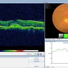 Choroidal Osteoma Plus CNV
Sep 2 2012 by Hamid Ahmadieh, MD
Color fundus photograph and OCT imaging of a 47-year-old man with a juxtafoveal CNV superimposed on a choroidal osteoma.
Photographer: Hamid Ahmadieh, Ophthalmic Research Center, Labbafinejad Medical Center
Imaging device: Topcon
Condition/keywords: choroidal neovascularization (CNV), choroidal osteoma, optical coherence tomography (OCT)
-
 Retinal Angiomatous Proliferation in Age-Related Macular Degeneration with Subretinal Neovascularization
Sep 24 2012 by James B. Soque, CRA, OCT-C, COA, FOPS
75-year-old white male with classic SRN with RAP. Lesion OD is active, and patient is receiving anti-VEGF treatment. Mid phase FA, 50 Deg, Mag 2x.
Photographer: James Soque, CRA, COA, Island Retina, Shirley, NY, USA
Imaging device: Topcon TRC 50 DX, OIS 5.0 MP Color, FA Camera, OIS Software
Condition/keywords: age-related macular degeneration (AMD), fundus autofluorescence (FAF), leakage, retinal angiomatous proliferation (RAP), subretinal neovascularization (SRNV)
-
 Punctate Inner Choroidopathy with CNV Treated with Bevacizumab # 6 of 7
Feb 28 2013 by Gregory R. Blaha, MD, PhD
Fundus photo following treatment with bevacizumab in a 31-year-old female with vision loss from a choroidal neovascular membrane (CNV) from punctate inner choroidopathy. The vision improved and was stable following a single injection.
Photographer: Gerard Gauthier, Spindel Eye Assoc., Derry, NH
Imaging device: Zeiss FF 450 Plus
Condition/keywords: bevacizumab, choroidal neovascularization (CNV), punctate inner choroidopathy (PIC)
-
 Ischemic BRVO with neovascularization
Aug 23 2012 by Gerardo Garcia-Aguirre, MD
Fluorescein angiogram of the temporal periphery showing wide areas of capillary nonperfusion and leakage secondary to neovascularization.
Photographer: Noemí Hernández, Asociación para Evitar la Ceguera en México
Condition/keywords: branch retinal vein occlusion (BRVO), capillary nonperfusion, neovascularization (NV)
-
 Pigment Epithelial Detachment late FA with small occult CNV
Jul 6 2012 by Tarek S. Hassan, MD, FASRS
72-year-old man with VA loss and metamorphopsia of 2 months duration. PED found, testing done to rule out CNV. Very suspicious for CNV in superonasal fovea/parafovea.
Condition/keywords: choroidal neovascularization (CNV), pigment epithelial detachment (PED)
-
 Proliferative Diabetic Retinopathy with Subhyaloid Hemorrhage
Oct 18 2012 by Suber S. Huang, MD, MBA, FASRS
43-year-old diabetic man with proliferative diabetic retinopahty, subhyaloid hemorrhage, ischemia, neovascuarization.
Photographer: Stacie Hrvatin
Condition/keywords: ischemia, retinal neovascularization, subhyaloid hemorrhage
-
 Proliferative Diabetic Retinopathy
Sep 17 2012 by Michael P. Kelly, FOPS
Retinal fundus photograph of a patient with PDR and NVD.
Photographer: Michael P. Kelly, FOPS Director, Duke Eye Labs, Duke University Hospital, Duke Eye Center
Imaging device: Topcon
Condition/keywords: blot hemorrhages, neovascularization of the disc (NVD)
-
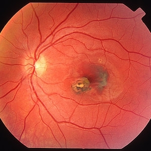
 Optic Nerve Head Drusen With Idiopathic CNV
Feb 17 2017 by Kristen Wagner
22-year-old female fundus photograph of a right eye with Optic Nerve Drusen with Idiopathic CNV.
Photographer: Kristen Wagner, COT, OSC Ophthalmic Photographer, Tennessee Retina, Nashville TN
Condition/keywords: choroidal neovascularization (CNV), drusen of optic disc, optic disc drusen
-
 Proliferative Diabetic Retinopathy - Neovascularization on the Disc
Aug 23 2012 by Gerardo Garcia-Aguirre, MD
Fluorescein angiogram, early phase, showing microaneurysms, wide areas of capillary nonperfusion, and leakage secondary to neovascularization on the disc.
Photographer: Noemí Hernández, Asociación para Evitar la Ceguera en México
Condition/keywords: microaneurysms, neovascularization of the disc (NVD)
-
 Ischemic BRVO with Neovascularization
Aug 23 2012 by Gerardo Garcia-Aguirre, MD
Fluorescein angiogram of the macula showing wide areas of capillary nonperfusion and leakage in the superotemporal quadrant.
Photographer: Noemí Hernández, Asociación para Evitar la Ceguera en México
Condition/keywords: branch retinal vein occlusion (BRVO), capillary nonperfusion, neovascularization (NV)
-
 Angle Neovascularization
Mar 21 2013 by Yusuke Oshima, MD, PhD
Angle neovascularization due to ischemic CRVO.
Photographer: Yusuke Oshima, MD, PhD, Osaka University Graduate School of Medicine
Condition/keywords: angle neovascularization, gonioscopy
-
 Secondary Choroidal Neovascularization Due to Toxoplasmosis
Feb 25 2013 by Henry J. Kaplan, MD
Left eye: secondary choroidal neovascularization and subretinal hemorrhage in a patient with old macular scar of toxoplasma.
Condition/keywords: choroidal neovascularization (CNV), toxoplasmosis, toxoplasmosis chorioretinitis
-
 Extra Macular CNV
Aug 24 2012 by John S. King, MD
P/C c mild VFD
Photographer: Kristin Konecki, OcuSight Eye Care Center, Rochester, NY
Condition/keywords: choroidal neovascularization (CNV)
-
 Angioid Streaks & CNV (Fig 3)
Aug 25 2012 by Hamid Ahmadieh, MD
Early phase ICG angiography imaging of a 53-year-old woman with a juxtafoveal CNV secondary to angioid streaks.
Photographer: Hamid Ahmadieh, Ophthalmic Research Center, Labbafinejad Medical Center
Imaging device: Heidelberg Spectralis
Condition/keywords: angioid streaks, choroidal neovascularization (CNV), indocyanine green (ICG) angiography
-
 Sea Fan Neovascularisation
Apr 27 2015 by Neha Goel, MS DNB FRCS (Glasg)
Fluorescein angiography of the left eye of a 40-year-old male.
Photographer: Neha Goel
Imaging device: Zeiss visucam
Condition/keywords: Eales disease, neovascularization elsewhere (NVE), vasculitis
-
 Angioid Streaks & CNV (Fig 5)
Sep 2 2012 by Hamid Ahmadieh, MD
OCT imaging of a 53-year-old woman with a juxtafoveal CNV secondary to angioid streaks.
Photographer: Hamid Ahmadieh, Ophthalmic Research Center, Labbafinejad Medical Center
Imaging device: Topcon
Condition/keywords: angioid streaks, choroidal neovascularization (CNV), optical coherence tomography (OCT)
 Loading…