Search results (1738 results)
-
 Rubeosis Iridis
Jul 21 2025 by Luai Abu-Ismail, MD
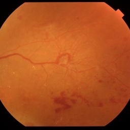
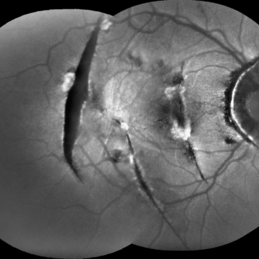
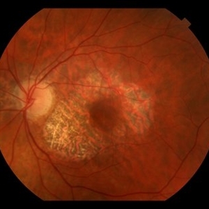
73y old female patient, known case of uncontrolled DM for more than 15y and chronic kidney disease. She had CRVO and complicated with 100 day neovascularization glaucoma.
Photographer: Luai Abu-Ismail
Imaging device: S23 Ultra
Condition/keywords: central retinal vein occlusion (CRVO), neovascular glaucoma, Rubeosis
-
 Venous Beading with Neovascularization
Jul 21 2025 by Moazzam Parvez
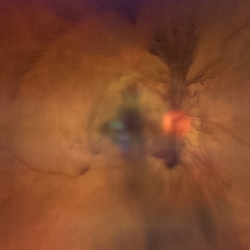
Fundus photograph of a 58 year old woman showing profuse venous beading and underlying neovascularization.
Photographer: Moazzam Parvez , Netralayam , Kolkata
Imaging device: Topcon Maestro 2
Condition/keywords: neovascularization (NV), periphery, Retina, venous beading
-
 Shooting Stars
Jul 9 2025 by Majda Hadziahmetovic, MD
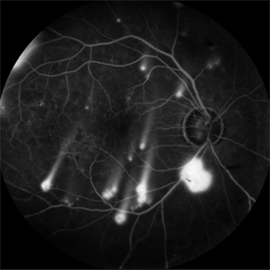
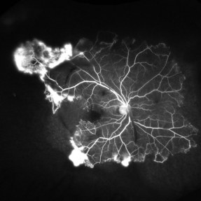
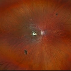
Fluorescein angiography image demonstrating multiple areas of neovascularization in a middle-aged male patient with long-standing diabetes.
Condition/keywords: proliferative diabetic retinopathy (PDR)
-
 Proliferative Sickle Cell Retinopathy
Jul 8 2025 by Niloofar Piri, MD
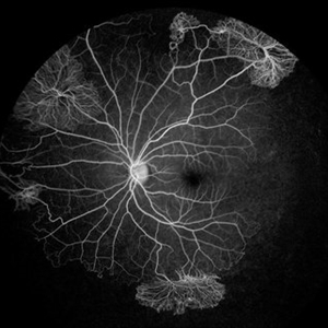
Mid AV phase fluorescein angiogram of a 13 yo AA male with SC disease demonstrating multiple classic sea fan neovascularization with peripheral capillary non perfusion (CNP). CNP is more obvious in this image involving the temporal retina and inferonasal retina.
Photographer: Stefan Raev, COT, Saint Louis University
Condition/keywords: Proliferative sickle cell retinopathy, proliferative sickle retinopathy, sickle cell retinopathy
-
 OCT Choroidal Rupture
Jun 26 2025 by Hector Gabriel Moreno Solano, MD, MHA
High-resolution OCT of the right eye shows a localized disruption of the retinal pigment epithelium (RPE)–Bruch’s membrane complex, consistent with a choroidal rupture. There is loss of the normal outer retinal architecture over the lesion, with focal elevation and irregularity of the underlying RPE. Hyperreflective material is noted at the level of the break, without associated subretinal fluid or signs of active choroidal neovascularization.
Photographer: Hector Gabriel Moreno Solano, Instituto Mexicano de Oftalmología “IMO I.A.P”
Imaging device: REVO
Condition/keywords: Choroidal Rupture, OCT
-
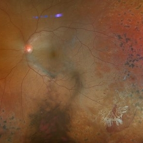
 Retinal Vasoproliferative Tumor
Jun 24 2025 by Marcelo Zas, MD PhD
We present a case of a 33-year-old male patient, who presented with decreased visual acuity in his right eye with 20/80, presenting a primary retinal vasoproliferative tumor in the lower temporal quadrant. The tumor is associated with serous retinal detachment, hard exudation, neovascularization and telangiectasias. Lipid exudates extend toward the macula, indicating macular involvement, which may contribute to decreased visual acuity. Oi was normal with 20/20 of BCVA. The patient was treated initially with IV anti-VEGF therapy and cryotherapy.
Photographer: Marcelo Zas MD PhD
Condition/keywords: RETINAL VASOPROLIFERATIVE TUMOR
-
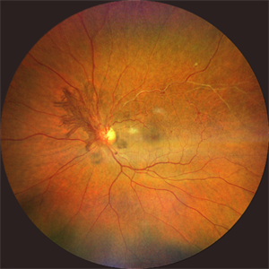
 Serpiginous Choroidopathy
Jun 23 2025 by César Adrián Gómez Valdivia, MD
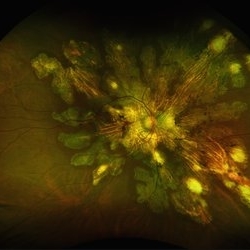
Fundus photograph of a 29 year-old female patient diagnosed with Serpiginous Choroidopathy. Finings were bilateral. The most common complication of SC is choroidal neovascularization affecting up to 35% of patients. Other reported complications are subretinal fibrosis, cystoid macular edema, branch vein occlusion, serous retinal detachment, optic disc neovascularization ,and anterior uveitis.
Photographer: @eyemissu2
Imaging device: TOPCON TRC-50DX
Condition/keywords: serpiginous choroiditis
-
 Serpiginous Choroidopathy
Jun 23 2025 by César Adrián Gómez Valdivia, MD
Fundus photograph of a 29 year-old female patient diagnosed with Serpiginous Choroidopathy. Finings were bilateral. The most common complication of SC is choroidal neovascularization affecting up to 35% of patients. Other reported complications are subretinal fibrosis, cystoid macular edema, branch vein occlusion, serous retinal detachment, optic disc neovascularization, and anterior uveitis.
Photographer: @eyemissu2
Imaging device: California ICG OPTOS
Condition/keywords: serpiginous choroiditis
-
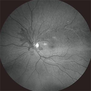
 Post-traumatic Choroidal Rupture
Jun 20 2025 by Alexander Babaev
FAF
Photographer: Babaev Alexander, Saint-Petersburg, medical clinic "Vision"
Imaging device: Carl Zeiss Visucam 500
Condition/keywords: choroidal neovascularization (CNV), Choroidal rupture
-
 Proliferative Sickle Retinopathy
Jun 13 2025 by Brandon I Fram, MD, BS
30 year-old with HbSC sickle retinopathy found to have profound retinal ischemia and florid peripheral neovascularization.
Imaging device: Fluorescein Angiography
Condition/keywords: proliferative sickle retinopathy, retinal ischemia, sea fan, sickle cell retinopathy
-
 Neovascularization of the Disc
Jun 3 2025 by Scott D Walter, MD, MSc, FASRS
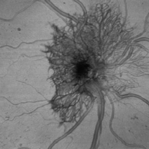
Near-infrared (NIR) en face OCT image showing neovascularization of the disc (NVD) in a patient with type II diabetes mellitus, complicated by proliferative diabetic retinopathy (PDR).
Imaging device: Heidelberg Spectralis
Condition/keywords: Diabetes, Heidelburg Spectralis, microaneurysms, Neovascularisation at the Disc (NVD), NEOVASCULARISATION OF DISC, OCT EN FACE, proliferative diabetic retinopathy (PDR)
-
 Proliferative Diabetic Retinopathy
May 29 2025 by KANWALJEET HARJOT MADAN, M.S. (Ophthalmology); FAICO (Vitreous - Retina)
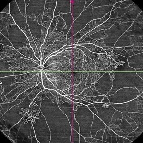
This is widefield optic coherence tomography angiography (WF-OCTA) picture of LE of a diabetic patient. This patient had Proliferative Diabetic Retinopathy and depicts large areas of capillary non perfusion with neovascularization elsewhere.
Photographer: Dr. Kanwaljeet Harjot Madan, Thind Eye Hospital, Jalandhar City (Punjab) INDIA.
Imaging device: Widefield Optic Coherence Tomography Angiography (WF-OCTA).
Condition/keywords: OCTA, proliferative diabetic retinopathy (PDR), ultra-wide field imaging
-
 Neovascularization of the Disc (NVD) - Red Free
Apr 28 2025 by Vishal Agrawal, MD, FRCS,FACS,FASRS
The red-free image enhances the visualization of the NVD, showing the fine neovascular fronds sprouting from the optic disc. Collateral vessels and vascular anastomosis are better appreciated.
Photographer: Dr Ayushi Gupta
Imaging device: Clarus 700
Condition/keywords: branch retinal vein occlusion (BRVO), collaterals
-
 Neovascularization of the Disc (NVD)
Apr 28 2025 by Vishal Agrawal, MD, FRCS,FACS,FASRS
Fundus image showing prominent neovascularization of the disc (NVD)- visible as fine, frond-like vascular proliferation extending from the disc surface.
Photographer: Dr Ayushi Gupta
Imaging device: Clarus 700
Condition/keywords: branch retinal vein occlusion (BRVO), NVD
-
 Neovascularisation Disc
Apr 4 2025 by Tejaswita Verma
Fundus photo of a 38 year-old Type 1 diabetic male with 6/18P vision in RE, presenting with TRD, NVD.
Photographer: DR. TEJASWITA VERMA
Imaging device: MIRANTE
Condition/keywords: neovascularization of the disc (NVD), TRD
-
 Actively Bleeding NVE
Apr 1 2025 by Jordyn Beckman
47 year old woman presented with actively bleeding NVE temporally on exam with complaints of foggy vision and floaters.
Photographer: Jordyn Beckman, Retina Consultants of Carolina, P.A.
Imaging device: Optos California
Condition/keywords: active bleeding, Elevated retinal neovascularization, vitreous hemorrhage
-
 Proliferative Diabetic Retinopathy S/P Pan Retinal Photocoagulation
Mar 4 2025 by Prithvi Chandrakanth
A 52-year-old female patient presented with complaints of diminishing vision, compounded by uncontrolled diabetes mellitus. Her Fundus examination revealed proliferative diabetic retinopathy, characterized by neovascularization of the disc and elsewhere, and sclerosed vessels. To address this, Pan Retinal Photocoagulation was performed, and the condition stabilized, halting the progression of the disease.
Photographer: DR PRITHVI CHANDRAKANTH, DR CHANDRAKANTH NETHRALAYA, KOZHIKODE, KERALA, INDIA
Imaging device: EIDON
Condition/keywords: Diabetic Retinopathy, Neovascularisation at the Disc (NVD), neovascularization of the disc (NVD), NVD, pan-retinal photocoagulation (PRP), PDR, PDR with NVE (periphery), PRP
-
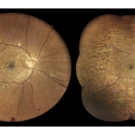
 MIDD (Maternally Inherited Diabetes and Deafness)
Feb 25 2025 by Virginia Gebhart
53 year old female with confirmed MIDD (genetic testing at Emory). Vision is stable with progressing GA but still central sparing OU. No evidence of choroidal neovascularization. Moderate myopia.
Photographer: Virginia Gebhart, Retina Consultants of Carolina
Imaging device: Topcon 50DX
Condition/keywords: geographic atrophy, Maternally inherited diabetes and deafness (MIDD), MIDD
-
 Diabetic Retinopathy
Feb 19 2025 by Martina Rubesova
Venous looping and beading are caused by severe retinal hypoxia and indicate an increased risk for progression to neovascularization.
Photographer: Ivan Kolin, lexum
Imaging device: Clarus Zeiss
Condition/keywords: diabetic retinopathy, venous beading
-
 Myopic CNVM
Jan 31 2025 by Thirumalesh Mochi Basavaraj, MD
Widefield image of a 26 year-old male patient with pathologic myopia with history of central scotoma with a sub macular bleed.
Photographer: Puttaswamy N K
Imaging device: Optos Daytona
Condition/keywords: myopic choroidal neovascularization (CNV), Myopic CNVM, pathologic myopia
-
 Eales Disease
Jan 31 2025 by Thirumalesh Mochi Basavaraj, MD
Ultra wide field image of a 24 year-old young healthy adult male with a visible sea fan neovascularization with partial PVD secondary to Scatter LASER photocoagulation with Vitreous and subhyaloid hemorrhage.
Photographer: Puttaswamy N K
Condition/keywords: Eales disease, Neovascularisation elsewhere (NVE), sea fan
-
 Eales Disease
Jan 31 2025 by Thirumalesh Mochi Basavaraj, MD
Ultra-wide field image of a 24 year old young healthy adult male with a visible sea fan neovascularization with partial PVD with vitreous and subhyaloid hemorrhage.
Photographer: Puttaswamy
Condition/keywords: Eales disease, sea fan, Ultra-wide field retinal imaging
-
 Proliferative Sickle Cell Retinopathy
Jan 27 2025 by Virginia Gebhart
61 year-old with proliferative sickle cell retinopathy s/p cryotherapy to peripheral fibrotic NV. Eye is stable with resolving exudates and maturing cryo scar. BCVA 20/40
Photographer: Virginia Gebhart, Retina Consultants of Carolina
Imaging device: Optos California
Condition/keywords: cryotherapy, fibrotic neovascularization, sickle cell retinopathy
-
 Raised NVD
Jan 9 2025 by Richa Chaudhary, Mbbs,ms
62 year old female, with h/o vein occlusion, presented with florid raised NVE , regressed well with intravitreal anti-VEGF.
Condition/keywords: neovascularization of the disc (NVD)
-
 Inactive Chorioretinal Scars
Dec 11 2024 by Virginia Gebhart
30 year old female with chorioretinal and macula scars. Appears post-infectious, most likely toxoplasmic. No active inflammatory changes or choroidal neovascularization. Will continue to monitor. Central vision limited by macula scar, BCVA 20/100
Photographer: Virginia Gebhart, Retina Consultants of Carolina
Imaging device: Optos California
Condition/keywords: chorioretinal scar, inactive toxoplasmosis
 Loading…