Search results (22 results)
-
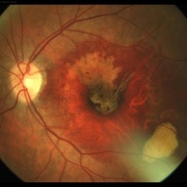
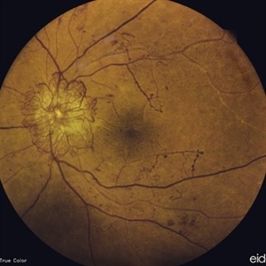
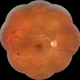
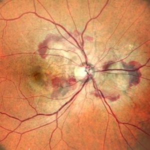 Right Eye Color Photo With Hemorrhages in Case of CNVM With Angioid Streaks
Nov 29 2024 by Anand Temkar
A 45 year old male came with chief complaint of blurring vision in right eyes since past 4 days. His vision is 6/12 in right eye and 6/9 in left eye. His vision was 14 mmHg in right eye and 16 mmHg in left eye. He was diagnosed with Angioid Streaks in both eyes about a year ago, then he developed choroidal neovascularization in his left eye 8 months ago, for which he received AntiVEGF injections x 3. Left eye is a stable eye now. Patient presented with right eye choroidal neovascularization in a case of Angioid Streaks on recent follow up. We have advised him right eye AntiVEGF injections x 3. In this image, the right eye color photo shows bleed from CNVM in case of angioid streaks.
Photographer: Dr.Anand Temkar- Retina Foundation, Ahmedabad
Imaging device: Mirante
Condition/keywords: Angioid Streaks, choroidal neovascular membrane (CNVM)
-
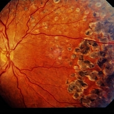
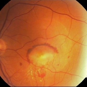
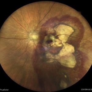 Subretinal Hemorrhage
Feb 28 2023 by Akansha Sharma
Color fundus photograph of an 84-year old male with subretinal hemorrhage associated with areas of scarring.
Photographer: Dr. Urmil Shah, Dr. Denish Patel, Dr. Akansha Sharma, Bharati Eye Hospital, Ahmedabad, Gujarat
Condition/keywords: choroidal neovascularization (CNV), subretinal hemorrhage
-
 Neovascular vessels
Sep 22 2022 by Filip Kecer
Multicolor widefield scan of a 16-year-old girl with a neovascularization from disc to vitreous space
Photographer: Filip Kecer, National Institute of Childrens Diseases
Imaging device: Spectralis, Heidelberg Engineering
Condition/keywords: neovascularization (NV), neovascularization at the disc, uveitis, vitreous
-
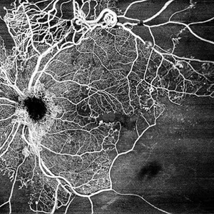 Central Retinal Vein Occlusion by OCT Angiography
Jun 13 2022 by JORGE SOBERANES
A 63 year old man with a central retinal vein oclussion. In the OCT angiogram we could observe retinal isquemia, neovascularization and arteriovenous shunts.
Photographer: Jorge I. Soberanes MD
Imaging device: PLEX Elite 9000, Zeiss
Condition/keywords: Central vein oclussion, neovascularization, OCT angiography, retina, Shunts
-
 RPE Tear After Anti-VEGF Injection
Mar 17 2021 by RAFAEL REIS PEREIRA, MD
Retinal pigment epithelium (RPE) tear is a rare devastating complication of age-related macular degeneration (AMD). The believed mechanism lies in an adherence of the neovascularization to the undersurface of the RPE creating a contractile force that increases after VEGF therapy and causes the tear.
Photographer: Rafael Reis, Retina Clinic, São Paulo
Condition/keywords: retinal pigment epithelium (RPE) contracture
-
 Sickle Cell Retinopathy
Feb 15 2021 by Kim Barrett
24-year-old female with Sickle Cell Retinopathy, stage 3. She confirms she has the trait as well as her grandmother, mother and a sibling. She has seafan neovascularization superotemporal OD. Current VA is 20/20. Photo is pre-PRP laser with areas of non-profusion temporally.
Photographer: Kim Barrett C.O.A. Retina Specialist of Michigan, Grand Rapids, MI
Imaging device: Optos California
Condition/keywords: neovascularization (NV), pan-retinal photocoagulation (PRP), sickle cell retinopathy, stage 3, trait
-
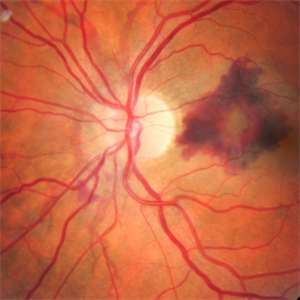
 Choroidal Neovascularization
May 27 2020 by Jamin S. Brown, MD
73-year-old female with CNV.
Photographer: Jeffrey Barker, Retina-Vitreous Surgeons of CNY
Condition/keywords: choroidal neovascularization (CNV)
-
 Branch Retinal Vein Occlusion with Acute on Chronic Subhyaloid Hemorrhage
Oct 24 2019 by Nichole Lewis
60-year-old male with a branch retinal vein occlusion and subhyaloid hemorrhage and retinal neovascularization. VA HM.
Photographer: Nichole Lewis
Condition/keywords: branch retinal vein occlusion (BRVO), retinal neovascularization, subhyaloid hemorrhage
-
 Subretinal Fibrosis (PPCNVM and POHS) OS
Sep 18 2019 by John S. King, MD
57-year-old white male with history of PPCNVM OS and POHS OU here for a routine visit. History of avastin in 2014, and stable since then. Va OS 20/20. PP scar with macular subretinal fibrosis. No heme or exudates. CR spot supero-nasally.
Photographer: Shelly Blair
Imaging device: Topcon 50
Condition/keywords: choroidal neovascular membrane (CNVM), ocular histoplasmosis syndrome (OHS), peripapillary choroidal neovascularization (PPCNVM), presumed ocular histoplasmosis syndrome (POHS)
-
 Proliferative Diabetic Retinopathy
Jul 9 2019 by Chinmayi Vyas
38-year-old type 1 diabetic female with gross neovascularization of disc.
Photographer: Sangeeta
Condition/keywords: neovascularization of the disc (NVD)
-
 Angioid Streaks
Apr 3 2019 by HECTOR LUIS VILLARROEL GUIZAR, MD, RETINA FELLOW
Retinal angiography composition of an 45-year-old man with angioid streaks and secondary choroidal neovascularization.
Photographer: HECTOR VILLARROEL, HOSPITAL DE LA LUZ, MEXICO CITY
Condition/keywords: angioid streaks
-
 ICG: Choroidal Aspergilloma With Secondary Choroidal Neovascularization and Exudative Retinal Detachment
Mar 21 2019 by Scott D Walter, MD, MSc, FASRS
Multimodal imaging of a transplant patient with disseminated Aspergillosis and vision loss in her left eye.
Condition/keywords: choroidal neovascular membrane (CNVM), choroidal neovascularization (CNV), exudative detachment, focal chorioretinitis, fungal endophthalmitis, granulomatous choroiditis
-
 Proliferative Diabetic Retinopathy (PDR)
Jul 4 2018 by Deepak Bhojwani, MS
Colour Fundus Photograph of a 66-year-old diabetic male with large fibro-vascular proliferative vessels causing subhayolid haemorrhage and tractional retinal detachment involving posterior pole.
Photographer: Deepak Bhojwani
Condition/keywords: diabetes, neovascularization (NV), subhyaloid hemorrhage, tractional retinal detachment
-
 Subhyaloid Hemorrhage With Flat Neovascular Vessels
Sep 26 2017 by Purva Patwari
60-year-old patient with uncontrolled diabetes.
Photographer: Dr Purva Patwari, Patwari Retina Clinic,Ahmedabad, India
Imaging device: Zeiss
Condition/keywords: flat neovascularization
-
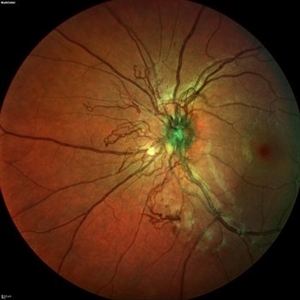
 Optic Nerve Head Drusen With Idiopathic CNV
Feb 17 2017 by Kristen Wagner
22-year-old female fundus photograph of a right eye with Optic Nerve Drusen with Idiopathic CNV.
Photographer: Kristen Wagner, COT, OSC Ophthalmic Photographer, Tennessee Retina, Nashville TN
Condition/keywords: choroidal neovascularization (CNV), drusen of optic disc, optic disc drusen
-
 Sickle Cell Neovascularization and Vitreous Hemorrhage
Oct 30 2015 by David Callanan, MD
Female patient, sickle cell neovascularization and vitreous hemorrhage; pre and post laser.
Condition/keywords: neovascularization (NV), sickle cell, vitreous hemorrhage
-
 RIP 2 FAF
Oct 7 2015 by Roberto Gallego-Pinazo, MD, PhD, DiSSO
Multicolor and autofluorescence sequence of a retinal pigment epithelium tear following intravitreal anti-VEGF injection.
Photographer: Rosa Dolz-Marco, University and Polytechnic Hospital La Fe, Valencia, Spain
Condition/keywords: age-related macular degeneration (AMD), autofluorescence imaging, choroidal neovascularization (CNV), multicolor, retinal pigment epithelium (RPE) tear
-
 Sea Fan Neovascularisation
Apr 27 2015 by Neha Goel, MS DNB FRCS (Glasg)
Fluorescein angiography of the left eye of a 40-year-old male.
Photographer: Neha Goel
Imaging device: Zeiss visucam
Condition/keywords: Eales disease, neovascularization elsewhere (NVE), vasculitis
-
 CNV Due to Angioid Streaks
Dec 6 2014 by Simon Kelly, MB, FRCSEd, FRCOphth, FEBO, DHC
Fundus photograph of left eye of 40-year-old female with recent onset of central distortion of left vision. Note the presence of angioid streaks radiating outwards from optic disc and macular bleeding. Treatment with intravitreal anti-VEGF injections was commenced.
Condition/keywords: angioid streaks, choroidal neovascular membrane (CNVM), choroidal neovascularization (CNV)
-
 Severe Neovascularization Secondary to Idiopathic Occlusive Retinal Vasculitis
Jan 17 2015 by Hamid Ahmadieh, MD
Wide- field color fundus photograph of the right eye of a 28-year-old woman with severe retinal neovascularization secondary to idiopatic occlusive retinal vasculitis.
Photographer: Solmaz Shahmohammad, Negah Eye Center, Tehran
Condition/keywords: color fundus photograph, neovascularization (NV), retinal vasculitis
-
 CNV Due to Chronic Central Serous Retinopathy
Apr 6 2014 by Ratimir Lazic, MD, PhD
A color fundus image of a 66-year-old male with previously diagnosed chronic CSR. Few weeks ago the patient noticed rapidly worsening of the visual acuity on the left eye. Subretinal hemorrhage with big PED and subretinal exudation can be noticed. The image presents baseline clinical picture of the left eye. The antiVEGF intravitreal treatment have been started.
Photographer: Marko Lukic, University Eye Clinic Svjetlost
Imaging device: Zeis Visucam Lite 2
Condition/keywords: central serous chorioretinopathy (CSCR), choroidal neovascularization (CNV), subretinal hemorrhage
-
 Proliferative Diabetic Retinopathy - Neovascularization on the Disc
Aug 23 2012 by Gerardo Garcia-Aguirre, MD
Fluorescein angiogram, early phase, showing microaneurysms, wide areas of capillary nonperfusion, and leakage secondary to neovascularization on the disc.
Photographer: Noemí Hernández, Asociación para Evitar la Ceguera en México
Condition/keywords: microaneurysms, neovascularization of the disc (NVD)
 Loading…