Search results (1738 results)
-
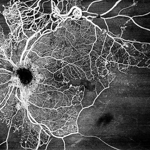
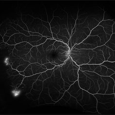
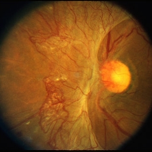
 Proliferative Diabetic Retinopathy
Sep 17 2012 by Michael P. Kelly, FOPS
Retinal fundus photograph of a patient with PDR and NVD.
Photographer: Michael P. Kelly, FOPS Director, Duke Eye Labs, Duke University Hospital, Duke Eye Center
Imaging device: Topcon
Condition/keywords: neovascularization of the disc (NVD)
-
 Central Retinal Vein Occlusion by OCT Angiography
Jun 13 2022 by JORGE SOBERANES
A 63 year old man with a central retinal vein oclussion. In the OCT angiogram we could observe retinal isquemia, neovascularization and arteriovenous shunts.
Photographer: Jorge I. Soberanes MD
Imaging device: PLEX Elite 9000, Zeiss
Condition/keywords: Central vein oclussion, neovascularization, OCT angiography, retina, Shunts
-
 Optic Nerve Head Drusen With Idiopathic CNV
Feb 17 2017 by Kristen Wagner
22-year-old female fundus photograph of a right eye with Optic Nerve Drusen with Idiopathic CNV.
Photographer: Kristen Wagner, COT, OSC Ophthalmic Photographer, Tennessee Retina, Nashville TN
Condition/keywords: choroidal neovascularization (CNV), drusen of optic disc, optic disc drusen
-
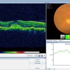
 Active CNVM on Angio OCT
Jul 11 2016 by Manish Nagpal, MD, FRCS (UK), FASRS
Angio OCT picture showing neovascularization corresponding to the area of CNVM.
Photographer: pooja barot
Condition/keywords: choroidal neovascular membrane (CNVM), optical coherence tomography (OCT)
-
 Cat Eye Syndrome
Feb 11 2020 by Sophia El Hamichi, MD
A 3-year-old female with cat eye syndrome including iris, chorioretinal and optic nerve colobomas. Note the CNV temporally to the optic nerve coloboma (blue arrows)
Photographer: Giselle De Oliveira, Bascom Palmer Eye Institute, Miami
Imaging device: RetCam
Condition/keywords: cat eye syndrome, chorioretinal coloboma, choroidal neovascularization (CNV), coloboma, coloboma of optic disc, optic nerve coloboma
-
 Choroidal Neovascularization
May 27 2020 by Jamin S. Brown, MD
73-year-old female with CNV.
Photographer: Jeffrey Barker, Retina-Vitreous Surgeons of CNY
Condition/keywords: choroidal neovascularization (CNV)
-
 NVI
NVI
Oct 24 2024 by Korey Starkey
Iris FA of a 74 year old male with neovascularization of the iris. Noted mild activity of NVI at the superior pupillary margin, recommending observation at time of visit.
Photographer: Korey Starkey
Imaging device: Heidelberg Spectralis
Condition/keywords: FA, Heidelburg Spectralis, Iris, iris fluorescein angiogram, neovascularization of iris (NVI), smokestack
-
 Proliferative Diabetic Retinopathy with Severe Ischemia
Nov 30 2023 by Gabriel Costa Andrade, PhD
Ultra-widefield fluorescein angiography of the right eye of a 47 year old woman with diabetes mellitus showing macular and nasal retinal capillary dropout and neovascularization of the disc and temporal vascular arcades.
Photographer: Gabriel Andrade
Imaging device: Optos California
Condition/keywords: Diabetic Retinopathy
-
 Proliferative Diabetic Retinopathy with Pre-retinal Hemorrhage
Jan 16 2018 by Olivia Rainey
Ultra-wide field pseudo-color image of an 57-year-old male with a large pre-retinal hemorrhage secondary to proliferative diabetic retinopathy affecting his left eye.
Photographer: Olivia Rainey
Imaging device: Optos California
Condition/keywords: color fundus photograph, diabetic mellitus, hemorrhage, left eye, neovascularization (NV), Optos, proliferative diabetic retinopathy (PDR), pseudocolor, ultra-wide field imaging
-
 Proliferative Diabetic Retinopathy with Subhyaloid Hemorrhage
Oct 18 2012 by Suber S. Huang, MD, MBA, FASRS
43-year-old diabetic man with proliferative diabetic retinopahty, subhyaloid hemorrhage, ischemia, neovascuarization.
Photographer: Stacie Hrvatin
Condition/keywords: ischemia, retinal neovascularization, subhyaloid hemorrhage
-
 Proliferative Sickle Cell Retinopathy
Jan 29 2021 by Olivia Rainey
Ultra-widefield fluorescein angiogram of a 24-year-old female with proliferative sickle cell retinopathy affecting her right eye. The physician's interpretation of the fluorescein shows seafan neovascularization superotemporally, AV anastomeses, and good peripheral laser. He performed scatter PRP OD on 12/2/2020 to nonperfusion in temporal far periphery. The patient's 12/2020 Hb electrophoresis came back showing Hb SC (rather than sickle cell trait). Patient was born at full term, but she reports that her mother used drugs while pregnant with the patient. The patient also mentioned that her niece has full sickle cell disease and her grandmother, mother, and sibling all have condition on the sickle cell spectrum.
Photographer: Olivia Rainey, OCT-C, COA
Imaging device: Optos California
Condition/keywords: fluorescein angiogram (FA), fluorescein leakage, neovascularization (NV), neovascularization elsewhere (NVE), Optos, sea fan, sickle cell retinopathy
-
 Proliferative Sickle Cell Retinopathy
Feb 1 2023 by Olivia Rainey
Ultra-widefield fluorescein angiography of a 25-year old male with Proliferative Sickle Cell Retinopathy affecting his right eye. Patient stated that he was born with Sickle disease (SC), and has yearly eye exams. He noted no vision concerns over the last year but has typically experienced sickle attacks about 1-2 per year. The physician noted that the fluorescein obtained showed peripheral nonperfusion affecting the patient's nasal and temporal retina as well as neovascularization affecting his left eye more than his right. He recommended pan retinal photocoagulation in his left eye for his temporal and nasal retina, as as well as his right eye following.
Photographer: Olivia Rainey, OCT-C, COA
Imaging device: Optos California
Condition/keywords: early phase, fluorescein angiogram (FA), fluorescein leakage, neovascularization (NV), non-perfusion, proliferative retinopathy, right eye, sickle cell retinopathy, ultra-wide field imaging, ultra-widefield image
-
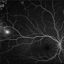
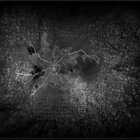
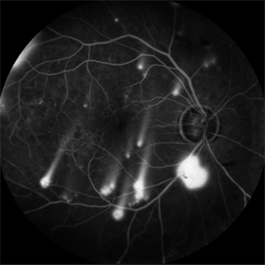 Shooting Stars
Jul 9 2025 by Majda Hadziahmetovic, MD
Fluorescein angiography image demonstrating multiple areas of neovascularization in a middle-aged male patient with long-standing diabetes.
Condition/keywords: proliferative diabetic retinopathy (PDR)
-
 Sickle Cell Retinopathy
Feb 15 2021 by Kim Barrett
24-year-old female with Sickle Cell Retinopathy, stage 3. She confirms she has the trait as well as her grandmother, mother and a sibling. She has seafan neovascularization superotemporal OD. Current VA is 20/20. Photo is pre-PRP laser with areas of non-profusion temporally.
Photographer: Kim Barrett C.O.A. Retina Specialist of Michigan, Grand Rapids, MI
Imaging device: Optos California
Condition/keywords: neovascularization (NV), pan-retinal photocoagulation (PRP), sickle cell retinopathy, stage 3, trait
-
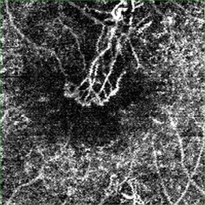
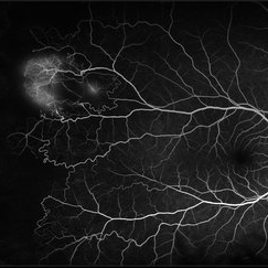
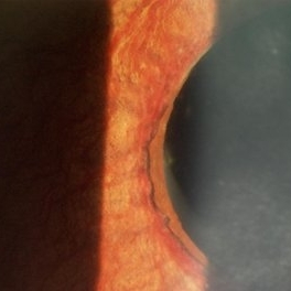
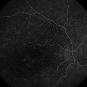
 "NVD Flower"
Oct 20 2023 by Daniel Davis, OCT-C
Infrared image of NVD (52F)
Imaging device: Heidelberg Spectralis
Condition/keywords: neovascularization of the disc (NVD)
-
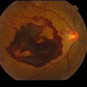
---thumb.jpg/image-square;max$300,300.ImageHandler) Active Choroidal Neovascularization With Subretinal Hemorrhage
Active Choroidal Neovascularization With Subretinal Hemorrhage
Nov 25 2013 by Maurice F. Rabb
Active choroidal neovascularization with subretinal hemorrhage.
Condition/keywords: choroidal neovascularization (CNV), subretinal hemorrhage
-
 Advanced Active PDR
Mar 29 2013 by Henry J. Kaplan, MD
Extensive NVD-FPD and NVE-FPE in a diabetic patient.
Condition/keywords: foveal photoreceptor defect, FPE, neovascularization (NV), neovascularization of the disc (NVD)
-
 Advanced Stage of Neovascular Glaucoma
Mar 21 2013 by Yusuke Oshima, MD, PhD
An 82-year-old man with a advanced stage of neovascular glaucoma. A slit-lamp photograph illustrates iris ectropion with prominent iris neovascularization.
Photographer: Yusuke Takada, Osaka University Graduate School of Medicine
Condition/keywords: neovascular glaucoma
-
 Angioid Streaks
Apr 3 2019 by HECTOR LUIS VILLARROEL GUIZAR, MD, RETINA FELLOW
Retinal angiography composition of an 45-year-old man with angioid streaks and secondary choroidal neovascularization.
Photographer: HECTOR VILLARROEL, HOSPITAL DE LA LUZ, MEXICO CITY
Condition/keywords: angioid streaks
-
 Central Retinal Vein Occlusion with Retinal Neovascularization
Jan 19 2022 by Olivia Rainey
Ultra-widefield fluorescein angiogram of a 56-year-old male with a Central Retinal Vein Occlusion with Retinal Neovascularization affecting his left eye. The patient presented on 1/19/2022 with scNLP vision in the left eye. The patient has good PRP, however areas of ischemia still remain untreated by laser. He also has severe neovascular glaucoma contributing to his poor vision.
Photographer: Olivia Rainey, OCT-C, COA
Imaging device: Optos California
Condition/keywords: central retinal vein occlusion (CRVO), FA early phase, fluorescein angiogram (FA), hemorrhage, ischemic CRVO, left eye, neovascular glaucoma, Optos, pan-retinal photocoagulation (PRP), retinal ischemia, retinal neovascularization, ultra-wide field imaging
-
 Choroidal Osteoma Plus CNV
Sep 2 2012 by Hamid Ahmadieh, MD
Color fundus photograph and OCT imaging of a 47-year-old man with a juxtafoveal CNV superimposed on a choroidal osteoma.
Photographer: Hamid Ahmadieh, Ophthalmic Research Center, Labbafinejad Medical Center
Imaging device: Topcon
Condition/keywords: choroidal neovascularization (CNV), choroidal osteoma, optical coherence tomography (OCT)
-
 CNV due to AMPPE
Oct 16 2012 by Ratimir Lazic, MD, PhD
Color fundus photography of a 58-year-old male. White dots with juxtafoveolar subretinal fluid can be seen. BCVA of that eye is 0.35.
Photographer: Marko Lukic, MD
Imaging device: Zeis Visucam Lite 2
Condition/keywords: acute posterior multifocal placoid pigment epitheliopathy (APMPPE), choroidal neovascularization (CNV)
-
 CNV due to AMPPE
Oct 16 2012 by Ratimir Lazic, MD, PhD
FAG of 58-year-old male. In early venous phase hyperflorescence of white dots (caused by window defect) can be seen. Leakage of dye in juxtafoveolar region.
Photographer: Marko Lukic, MD
Imaging device: Zeis Visucam Lite 2
Condition/keywords: acute posterior multifocal placoid pigment epitheliopathy (APMPPE), choroidal neovascularization (CNV)
-
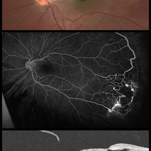 Coats Disease - Fundus Image, Angiography and OCT
Feb 3 2021 by Gabriel Costa Andrade, PhD
Fundus image, angiography and OCT of a 12-year-old boy with coats disease, pheripheral neovascularization and macular hole.
Photographer: Dr Gabriel Andrade, Dr André Maia
Condition/keywords: Coats' disease
-
 Corneal Abnormal Blood Vessels
Jul 14 2013 by Jason S. Calhoun
Corneal neovascularization, abnormal blood vessels growing on the epithelium.
Photographer: Jason S. Calhoun, Department of Ophthalmology, Mayo Clinic Jacksonville, Florida
Imaging device: TOPCON D-90 SL NIKON CAMERA
Condition/keywords: cornea
 Loading…