Search results (73 results)
-
 Diabetic Macular Edema, Proliferative Diabetic Retinopathy, Neovascularization Elsewhere, DME, PDR, NVE
Apr 1 2013 by James B. Soque, CRA, OCT-C, COA, FOPS
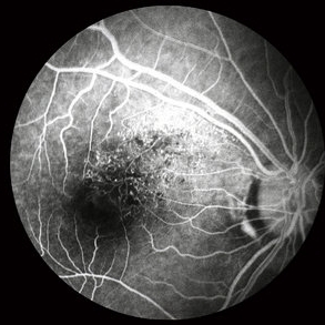
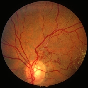
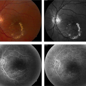
39-year-old white female and long standing diabetis, c/o new peripheral symptoms of left eye. FA OS reveals diabetic macular edema, microaneurysms, and neovasculaization elsewhere. Fluorescein Angogram, Early Phase, 50 Deg, 2x Mag.
Photographer: James B Soque, CRA, COA
Imaging device: Topcon TRC 50DX with MERGE software, OIS 10.6.45
Condition/keywords: diabetic macular edema, neovascularization (NV), proliferative diabetic retinopathy (PDR)
-
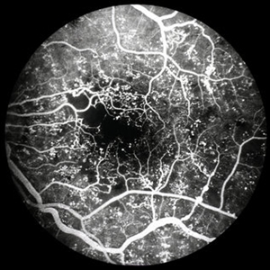
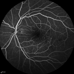
 Proliferative Diabetic Retinopathy - Neovascularization on the Disc
Aug 23 2012 by Gerardo Garcia-Aguirre, MD
Fluorescein angiogram, early phase, showing microaneurysms, wide areas of capillary nonperfusion, and leakage secondary to neovascularization on the disc.
Photographer: Noemí Hernández, Asociación para Evitar la Ceguera en México
Condition/keywords: microaneurysms, neovascularization of the disc (NVD)
-
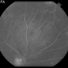
 BRVO FA, Early Phase
Oct 1 2012 by Jeffrey G. Gross, MD, FASRS
BRVO-FA early phase.
Condition/keywords: branch retinal vein occlusion (BRVO), capillary nonperfusion, early phase, microaneurysms
-
---thumb.jpg/image-square;max$300,300.ImageHandler) Peripheral retinal nonperfusion, capillary abnormalities, retinal microaneurysms, and intraretinal hemorrhage
Peripheral retinal nonperfusion, capillary abnormalities, retinal microaneurysms, and intraretinal hemorrhage
Feb 15 2013 by From the Collections of Thomas M. Aaberg, MD and Thomas M. Aaberg Jr., MD
Color fundus photograph showing peripheral retinal nonperfusion, capillary abnormalities, retinal microaneurysms, and intraretinal hemorrhage.
Condition/keywords: peripheral retinal nonperfusion, proliferative retinopathy
-
 PDR with Foveal Ischemia and FAZ Enlargement
Oct 8 2012 by Jeffrey G. Gross, MD, FASRS
PDR with foveal ischemia, and FAZ enlargement, multiple microaneurysms, FA, early phase.
Condition/keywords: early phase, enlarged foveal avascular zone, foveal ischemia, microaneurysms
-
 Proliferative Diabetic Retinopathy
Aug 23 2012 by Gerardo Garcia-Aguirre, MD
Fluorescein angiogram of a left eye of a 45 year-old patient with proliferative diabetic retinopathy. Small hyperfluorescent dots are observed (microaneurysms), as well as blockage from a subhyaloid hemorrhage. In the inferonasal area two areas of leakage secondary to neovascularization are observed.
Photographer: Noemí Hernández, Asociación para Evitar la Ceguera en México
Condition/keywords: microaneurysms, neovascularization (NV), subhyaloid hemorrhage
-
 Proliferative Diabetic Retinopathy
Oct 15 2012 by Susanna S. Park, MD, PhD
Fluorescein angiogram of the left eye of a 65 year old woman with diabetes mellitus showing nasal peripheral retinal capillary dropout and neovascularization of the disc. Scattered retinal microaneurysms are also noted
Photographer: Ellen Redenbo, University of California Davis Eye Center
Imaging device: Optos
Condition/keywords: proliferative diabetic retinopathy (PDR)
-
 Proliferative Diabetic Retinopathy - Neovascularization on the Disc
Aug 23 2012 by Gerardo Garcia-Aguirre, MD
Fluorescein angiogram, late phase, showing microaneurysms, wide areas of capillary non-perfusion, and leakage secondary to neovascularization on the disc.
Photographer: Noemí Hernández, Asociación para Evitar la Ceguera en México
Condition/keywords: microaneurysms, neovascularization of the disc (NVD)
-
 Proliferative Diabetic Retinopathy
Aug 23 2012 by Gerardo Garcia-Aguirre, MD
Fluorescein angiogram of a left eye of a 45-year-old patient with proliferative diabetic retinopathy. Small hyperfluorescent dots are observed (microaneurysms), as well as blockage from a subhyaloid hemorrhage.
Photographer: Noemí Hernández, Asociación para Evitar la Ceguera en México
Condition/keywords: microaneurysms, subhyaloid hemorrhage
-
 PDR NVD NVE
Jul 21 2014 by Susanna S. Park, MD, PhD
Mid-transit view fluorecein angiogram of the right eye of a 59-year-old diabetic woman with minimal peripheral fundus changes suggestive of diabetic retinopathy showing diffuse leakage of the disc and focal leakage in the peripheral retina from neovascularization. Peripheral retinal ischemia and leaking retinal microaneurysms are also seen.
Photographer: Karishma Chandra, University of California Davis Eye Center
Condition/keywords: fluorescein leakage, neovascularization of the disc (NVD), proliferative diabetic retinopathy (PDR)
-
---thumb.jpg/image-square;max$300,300.ImageHandler) Peripheral retinal nonperfusion, venous beading and dilatation, retinal microaneurysms, and intraretinal hemorrhage
Peripheral retinal nonperfusion, venous beading and dilatation, retinal microaneurysms, and intraretinal hemorrhage
Feb 15 2013 by From the Collections of Thomas M. Aaberg, MD and Thomas M. Aaberg Jr., MD
Color fundus photograph corresponding to slide titled "staining of retinal vessels, leakage from peripheral retinal neovascularization and peripheral nonperfusion." Shows peripheral retinal nonperfusion, venous beading and dilatation, retinal microaneurysms, and intraretinal hemorrhage.
Condition/keywords: peripheral retinal nonperfusion, proliferative retinopathy, retinal neovascularization
-
 NPDR
NPDR
Mar 29 2013 by Henry J. Kaplan, MD
Multiple microaneurysms visible as small round dot lesions.
Condition/keywords: nonproliferative diabetic retinopathy
-
 BRVO - longstanding
Jan 11 2013 by Alex P. Hunyor, MD
Longstanding branch retinal vein obstruction with collaterals, telangiectasia, microaneurysms and exudates - color image 1.
Condition/keywords: branch retinal vein occlusion (BRVO), collaterals
-
 Old Macular BRVO
Oct 10 2015 by Hamid Ahmadieh, MD
Mid -venous phase FA of the left eye of a 58-year-old woman with the history of macular BRVO. Note microaneurysms, collaterals , tortuosity of venules and an enlarged irregular FAZ compatible with macular ischemia.
Photographer: Nayereh Hadipour, Negah Eye Center, Tehran, Iran
Condition/keywords: collaterals, macular branch retinal vein occlusion (BRVO), macular ischemia, microaneurysms
-
 BRVO - longstanding
Jan 11 2013 by Alex P. Hunyor, MD
Longstanding branch retinal vein obstruction with collaterals, telangiectasia, microaneurysms and exudates - color image 2.
Condition/keywords: branch retinal vein occlusion (BRVO)
-
---thumb.jpg/image-square;max$300,300.ImageHandler) Hypertensive Retinopathy
Hypertensive Retinopathy
Oct 15 2013 by Maurice F. Rabb
The patient is a 61 year old female who first noted decreased vision in OS one year ago. The right eye is asymptomatic. The patient has systemic hypertension. There is a questionable history of angina. She has low back pain and leg pains of a non-specific nature. Uncorrected vision is OD was 20/25-, not improvable, and in OS 20/400, not improvable. Blood pressure recorded in the office was 230/105. There was mild nuclear sclerosis. Both fundi are characterized by multiple vascular abnormalities consistent with nerve fibers infarcts, hemorrhages (both deep and superficial), and occasional microaneurysms. In OS, there is a large subretinal and preretinal hemorrhage in the macula. This is surrounded by a wreath of outer retinal exudates.
Condition/keywords: hypertensive retinopathy
-
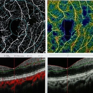 OCTA of Diabetic Retinopathy
Mar 13 2017 by Hashim Ali Khan, OD, FAAO
Optical coherence tomographic angiography showing capillary dropout and microaneurysms.
Imaging device: Angiovue
Condition/keywords: capillary dropouts, capillary nonperfusion, diabetic maculopathy, optical coherence tomography (OCT), retinal microaneurysms
-
 BRVO, FA, Hemorrhage, Diabetic
Mar 13 2014 by James B. Soque, CRA, OCT-C, COA, FOPS
51-year-old white male, diabetes, and with BRVO left eye, early phase 36 seconds. Flame heme from ON, showing microaneurysims, and fine capillary detail of this FA.
Photographer: James B Soque, CRA COA
Imaging device: Topcon TRC 50DX with MERGE software
Condition/keywords: branch retinal vein occlusion (BRVO), diabetes, FA early phase, microaneurysms
-
 Vascular Anormalities
Jan 6 2016 by Andrea Arriola-Lopez, MD MSc
77-year-old man. Decrease of visual acuity OS. VA 20/30 IOP 14mmHg. Fundus examination findings: Hard exudates, microaneurysms near to fovea. OCT shows IRF. Late leakage on FA.
Photographer: Andrea Elizabeth Arriola-Lopez, MSc MD
Condition/keywords: abnormal retinal vessel, aneurysm, hard exudates, vascular anomaly
-
---thumb.jpg/image-square;max$300,300.ImageHandler) peripheral retinal nonperfusion, capillary abnormalities, leaking retinal microaneurysms, and blocked fluorescence
peripheral retinal nonperfusion, capillary abnormalities, leaking retinal microaneurysms, and blocked fluorescence
Feb 15 2013 by From the Collections of Thomas M. Aaberg, MD and Thomas M. Aaberg Jr., MD
Mid-phase fluorescein angiograph showing peripheral retinal nonperfusion, capillary abnormalities, leaking retinal microaneurysms, and blocked fluorescence from intraretinal hemorrhage.
Condition/keywords: peripheral retinal nonperfusion, proliferative retinopathy
-
 Fluorescein Angiogram - Tortuous Vessels of DME Right Eye
Dec 10 2015 by James B. Soque, CRA, OCT-C, COA, FOPS
Early fluorescein angiogram of diabetic macular edema and tortuous vessels in the superior macula of the right eye.
Photographer: James B Soque, CRA, COA
Imaging device: Top[con TRC-50 DX with MERGE Winstation V 11.2.0
Condition/keywords: diabetes, diabetic macular edema, microaneurysms, microangiopathy, tortuous vessels
-
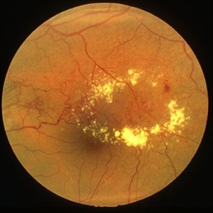
 Diabetic Macular Edema - Leaking Microaneurysms
Oct 3 2013 by Gerardo Garcia-Aguirre, MD
Diabetic macular edema - leaking microaneurysms.
Condition/keywords: diabetic macular edema, diabetic retinopathy circinate
-
 Macroaneurysm
Jan 13 2014 by David Callanan, MD
Macroaneurysm, HM 20/60 DM with microaneurysms in other eye, 50-year-old male.
Condition/keywords: macroaneurysm
-
---thumb.jpg/image-square;max$300,300.ImageHandler) Binder3 P12 Slide82
Binder3 P12 Slide82
Feb 15 2013 by From the Collections of Thomas M. Aaberg, MD and Thomas M. Aaberg Jr., MD
Color fundus photograph showing peripheral retinal nonperfusion, retinal neovascularization elsewhere (NVE), venous beading and dilatation, retinal microaneurysms, and intraretinal hemorrhage.
Condition/keywords: peripheral retinal nonperfusion, proliferative retinopathy, retinal neovascularization
-
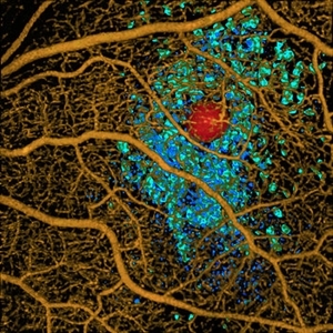 Volume Rendering Structural and Angiographic Optical Coherence Tomography Angiography Image of a Retinal Capillary Microaneurysm, A Newly Described Entity.
May 21 2019 by Richard F. Spaide, MD
This is a newly described entity in which patients develop solitary aneurysms that are much larger than typical microaneurysms and they are supplied by capillaries. The aneurysm is shown in red. The associated macular edema produced cystoid spaces in Henle’s fiber layer, rendered as teal and in the inner nuclear layer as blue.
Photographer: Richard F. Spaide, MD
Condition/keywords: aneurysm, optical coherence tomography (OCT), volume rendering
 Loading…