Search results (73 results)
-
 Diabetic Retinopathy
Jun 4 2025 by Paulina Araujo
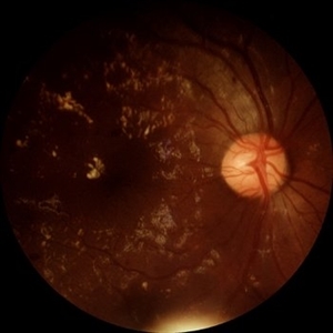
The 55-degree central fundus photograph of the right eye demonstrates numerous hard exudates, dot intraretinal hemorrhages, and microaneurysms.
Photographer: Paulina D.Araujo Martínez, Asociación para Evitar la Ceguera en México I.A.P., Hospital Dr Luis Sánchez Bulnes.
Condition/keywords: diabetic retinopathy
-
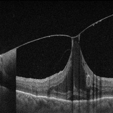
 Tractional Retinal Detachment
Jun 4 2025 by Paulina Araujo
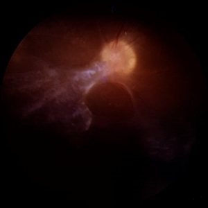
The 55-degree central fundus photograph of the right eye reveals a thickened and opacified hyaloid exerting traction on the optic disc and posterior pole of the retina, along with hard exudates and microaneurysms consistent with advanced proliferative diabetic retinopathy.
Photographer: Paulina D.Araujo Martínez, Asociación para Evitar la Ceguera en México I.A.P., Hospital Dr Luis Sánchez Bulnes.
Condition/keywords: tractional retinal detachment
-
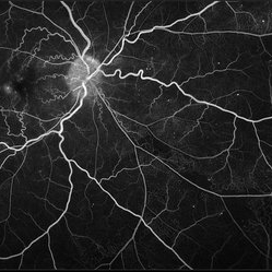
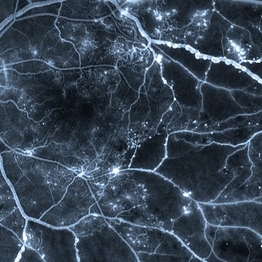
 Neovascularization of the Disc
Jun 3 2025 by Scott D Walter, MD, MSc, FASRS
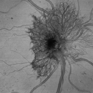
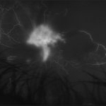
Near-infrared (NIR) en face OCT image showing neovascularization of the disc (NVD) in a patient with type II diabetes mellitus, complicated by proliferative diabetic retinopathy (PDR).
Imaging device: Heidelberg Spectralis
Condition/keywords: Diabetes, Heidelburg Spectralis, microaneurysms, Neovascularisation at the Disc (NVD), NEOVASCULARISATION OF DISC, OCT EN FACE, proliferative diabetic retinopathy (PDR)
-
 Retinal Microaneurysms & Dot/Blot Hemes Autofluorescence OS
May 12 2025 by Briana Hernandez
OS Autofluorescence Optos Image of Retinal Microaneurysms & Dot/Blot Hemes in 91-year-old female BRVO patient.
Photographer: Briana Hernandez, Hilton Head Retina Institute
Imaging device: Optos
Condition/keywords: Autoflourescence, branch retinal vein occlusion (BRVO)
-
 Retinal Microaneurysms & Dot/Blot Hemes Fundus Photo OS
May 12 2025 by Briana Hernandez
OS Optos Fundus Photo of Retinal Microaneurysms & Dot/Blot Hemes in 91-year-old female BRVO patient.
Photographer: Briana Hernandez, Hilton Head Retina Institute
Imaging device: Optos
Condition/keywords: macular
-
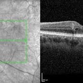
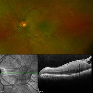
 Vitreomacular Adhesion Showcasing a Microaneurysm and a Subhyaloid Hemorrhage
Jan 3 2025 by Drew Mitchell
A vertical OCT 1 line raster scan positioned slightly inferomacula to document the subhyaloid hemorrhage. Hyper reflective Oval indicating Microaneurysm.
Photographer: Drew Mitchell, OCT-C
Imaging device: Zeiss Cirrus 5000
Condition/keywords: diabetic macular edema, microaneurysms, retinal microaneurysms, subhyaloid hemorrhage, subretinal fluid, vitreomacular adhesion, vitreomacular traction (VMT)
-
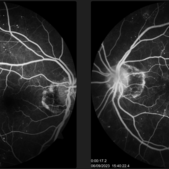
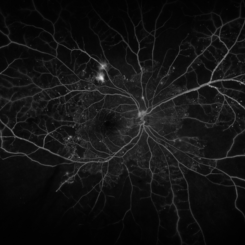
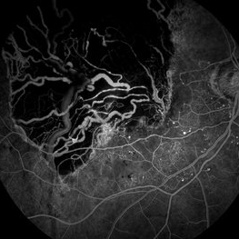
 Chronic CRVO
Dec 12 2024 by Korey Starkey
Fluorescein Angiography of a 62 year-old man with chronic central retinal vein occlusion. Vision is 20/200.
Photographer: Korey Starkey
Imaging device: Optos
Condition/keywords: capillary nonperfusion, central retinal vein occlusion (CRVO), FLUORESCEIN ANGIOGRAPHY, ischemia, microaneurysms, Optos
-
 Diabetic Retinopathy
Nov 20 2024 by Korey Starkey
64 year old female being monitored for moderate-severe diabetic retinopathy.
Photographer: Korey Starkey
Condition/keywords: capillary nonperfusion, FA, FLUORESCEIN ANGIOGRAPHY, microaneurysms, nonproliferative diabetic retinopathy, Optos, OPTOS CALIFORNIA, tortuous vessels
-
 Proliferative Diabetic Retinopathy with Neovascularization
Nov 15 2024 by Júlio Andrade
Fundus photograph of a patient with proliferative diabetic retinopathy presenting with extensive neovascularization, microaneurysms, and evidence of fluid leakage. Key findings include the presence of new blood vessel formation, indicative of advanced retinal ischemia and disease progression.
Photographer: úlio Andrade, Retina Instituto, Belo Horizonte
Imaging device: Zeiss Clarus 700
Condition/keywords: diabetic blindness, ischaemic diabetic maculopathy, Neovascularisation elsewhere (NVE), NEOVASCULARISATION OF DISC, Proliferative Diabetic retinopathy
-
 Branch Retinal Vein Occlusion
Aug 22 2024 by Virginia Gebhart
Fluorescein angiogram of branch retinal vein occlusion in 75 year old female. Scattered microaneurysms with late CME and persistent SRF. Pt will consider laser treatment but is hesitant for injections at this time due to possible side effects.
Photographer: Virginia Gebhart
Imaging device: Optos California
Condition/keywords: branch retinal vein occlusion (BRVO), BRVO, cystoid macular edema (CME), FA, FA late phase, fluorescein angiogram (FA), macular edema, microaneurysms, retinal microaneurysms
-
 Macular Telangiectasia Type 2 Fluorescein Angiography
Mar 29 2024 by Lucy V Cobbs, M.D.
Fluorescein angiography of the left eye of a 45-year-old African American female with MacTel type 2 and Type 2 Diabetes. This angiogram demonstrates peripheral microaneurysms characteristic of mild non proliferative diabetic retinopathy and temporal foveal leakage with telangiectatic macular capillaries classic for MacTel type 2. There is a well-established association between the two conditions.
Condition/keywords: Mac Tel type 2
-
 The Starry Sky
Mar 12 2024 by MEENAL SONI
57 year old female with DOV in BE, known diabetic. OU Multiple microaneurysms and haemorrhages at macula with diabetic macular oedema.
Photographer: Dr. Meenal Soni, Fellow VR, ASG eye Hospital Jodhpur
Imaging device: ZEISS Visucam 400
Condition/keywords: diabetic macular edema, Diabetic Retinopathy
-
 Microaneurysm With Intraretinal Fluid on Optical Coherence Tomography
Feb 22 2024 by Nikhil K Bommakanti, MD
Microaneurysm with associated intraretinal fluid on optical coherence tomography in mild nonproliferative diabetic retinopathy.
Condition/keywords: microaneurysm, retinal microaneurysms
-
 Idiophatic retinal vasculitis
Jul 9 2023 by Luiz A Zago, PhD
Mid phase angiography of a 45 woman with a idiophatic vasculitis. She is been followed in the last 10 years. No sistemic association was found beside face rosacea, This case was first described in Diagnostic and Therapeutic Challenges - Retina Journal
Photographer: Luiz Zago, PhD.
Imaging device: Topcon 50IX
Condition/keywords: anomalous foveal avascular zone, Choroidal Folds, intraretinal microvascular abnormalities, microaneurysms, optic neuritis, Vaculitis
-
 Venous Beading
Apr 30 2021 by Shivani Reddy, MD
This is a fluorescein angiogram image capturing a beautiful example of different stages of venous beading in diabetic retinopathy all in one frame. This patient also has various microangiopathic findings including microaneurysms, venous loops and capillary dropout. This patient is a 41 y/o male with a history of type 1 diabetes, presenting for his first eye exam in years.
Imaging device: Optos FA
Condition/keywords: capillary dropouts, nonproliferative diabetic retinopathy, proliferative diabetic retinopathy (PDR), retinal ischemia, venous beading
-
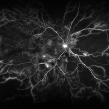
 PDR with Ischemia
Jul 7 2020 by Stephanie Burke
Early frame of a 45-year-old male with Type II diabetes.
Photographer: Stephanie Burke, CRA, OCT-C
Condition/keywords: FA early phase, ischemia, microaneurysms, neovascularization (NV), proliferative diabetic retinopathy (PDR), ultra-wide field imaging, venous beading
-
 Proliferative Diabetic Retinopathy
Apr 29 2020 by Stephanie Burke
Ultra-wide FA image of a 58-year-old man with DM.
Photographer: Stephanie Burke, CRA, OCT-C
Condition/keywords: ischemia, leakage, microaneurysms, neovascularization (NV), proliferative diabetic retinopathy (PDR), ultra-wide field imaging
-
 Macular Pucker
Jan 7 2020 by RAFAEL REIS PEREIRA, MD
A clinical grading system was proposed by Gass in 1987 describe the different stages of the epiretinal membrane. Grade 2 Macular pucker consists of a thick fibroglial membrane that contracts and produces obscuration of underlying vessels and marked full-thickness retinal distortion. Sometimes associated with cotton-wool spots, exudates, blot hemorrhages, microaneurysms, and cystoid macular edema.
Photographer: Rafael Reis, Retina Clinic - Brazil
Condition/keywords: macular pucker
-
 Retinal Macroaneurysm
Dec 17 2019 by Jonathan C. Tsui, MD
A 72-year-old female with uncontrolled hypertension presented with several spots in her vision. Fundus photography demonstrated a retinal macroaneurysm hemorrhage with subretinal fluid and intraretinal heme. Several microaneurysms are present adjacent to an anomalous vein which suggests a possible secondary venous occlusion.
Condition/keywords: retinal macroaneurysm
-
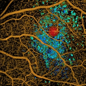 Volume Rendering Structural and Angiographic Optical Coherence Tomography Angiography Image of a Retinal Capillary Microaneurysm, A Newly Described Entity.
May 21 2019 by Richard F. Spaide, MD
This is a newly described entity in which patients develop solitary aneurysms that are much larger than typical microaneurysms and they are supplied by capillaries. The aneurysm is shown in red. The associated macular edema produced cystoid spaces in Henle’s fiber layer, rendered as teal and in the inner nuclear layer as blue.
Photographer: Richard F. Spaide, MD
Condition/keywords: aneurysm, optical coherence tomography (OCT), volume rendering
-
 Choroidal Melanoma - Stable, Fluorescein Angiogram, Early Phase
Mar 13 2019 by James B. Soque, CRA, OCT-C, COA, FOPS
Early FA, right eye, with choroidal melanoma-stable, and a few tiny microaneurysms showing leakage in re-circulation phase.
Photographer: James Soque, CRA, OCT-C, FOPS
Imaging device: Topcon TRC-50DX with MERGE Eye Station software
Condition/keywords: FA early phase, fluorescein angiogram (FA), MERGE, microaneurysms
-
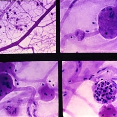 Slide 9-15
Feb 26 2019 by Lancaster Course in Ophthalmology
Trypsin digest retinal preparation showing diabetic microaneurysms. Some microaneurysms are relatively acellular (upper right and lower left), and others are filled with neutrophils (lower right).
Condition/keywords: microaneurysms, neutrophils, trypsin digestion
-
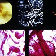 Slide 9-6
Feb 25 2019 by Lancaster Course in Ophthalmology
Coats' disease. An exudative detachment of the retina is shown with typical yellowish material and associated vascular abnormalities and some retinal hemorrhage (upper left). Fluorescein angiography shows capillary drop-out of the macroaneurysms and dilated telangiectatic vessels. Venous saccular dilatations also may be present (upper right). With trypsin digestion the background capillaries are acellular, and large, thin-walled telangiectatic vessels (lower left) and microaneurysms are present (lower right).
Condition/keywords: Coats' disease, macroaneurysm, telangiectatic vessels, trypsin digestion
-
 Tractional vs Combined Tractional/Rhegmatogenous Retinal Detachment with Active Neovascularization OS
Jun 1 2018 by Hosam Attia, MD
47-year-old African American, with history of diabetes mellitus of unknown duration and control, was referred for initial evaluation for conjunctival laceration in his left eye, following accidental finger nail injury, 6 days prior to presentation. - On exam, his vision was 20/50 OD and Bare HM/ LP OS. - Fundus color photos OD: No significant pathology, aside from attenuated vasculature OS: Chronic, Mac-Off, almost closed funnel tractional vs combined tractional/rhegmatogenous retinal detachment with large neovascularization (NVE) superiorly, detached ghost vessels, mild fresh vitreous hemorrhage, sub-retinal bands and inferior white vitreous debris from old hemorrhage (not shown) - FA OD: No significant pathology aside from possible mild capillary non-perfusion in the extreme periphery, attenuated vasculature and possible tiny microaneurysms, nasally. OS: Extensive, wide spread capillary non- perfusion (correlate w/ detached Ghost vessels on color photos), and leakage from the NVE. - B/L Carotid Duplex was recommended due to the striking asymmetry in pathology with unknown medical history, diabetes duration and control, etc (even in absence of any signs suggestive of possible ocular ischemic syndrome OD)
Imaging device: Optos California
Condition/keywords: combined retinal detachment, tractional retinal detachment
-
 Tractional vs Combined Tractional/Rhegmatogenous Retinal Detachment with Active Neovascularization OS
Jun 1 2018 by Hosam Attia, MD
47-year-old African American, with history of diabetes mellitus of unknown duration and control, was referred for initial evaluation for conjunctival laceration in his left eye, following accidental finger nail injury, 6 days prior to presentation. - On exam, his vision was 20/50 OD and Bare HM/ LP OS. - Fundus color photos OD: No significant pathology, aside from attenuated vasculature OS: Chronic, Mac-Off, almost closed funnel Tractional vs Combined Tractional/Rhegmatogenous Retinal Detachment with large neovascularization (NVE) superiorly, detached ghost vessels, mild fresh vitreous hemorrhage, sub-retinal bands and inferior white vitreous debris from old hemorrhage (Not shown) - FA OD: No significant pathology aside from possible mild capillary non-perfusion in the extreme periphery, attenuated vasculature and possible tiny microaneurysms, nasally. OS: Extensive, wide spread capillary non- perfusion (correlate w/ detached Ghost vessels on color photos), and leakage from the NVE. - B/L Carotid Duplex was recommended due to the striking asymmetry in pathology with unknown medical history, diabetes duration and control, etc (even in absence of any signs suggestive of possible ocular ischaemic syndrome OD)
Imaging device: Optos California
Condition/keywords: combined retinal detachment, tractional retinal detachment
 Loading…