Search results (73 results)
-
 Venous Beading
Apr 30 2021 by Shivani Reddy, MD
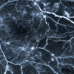
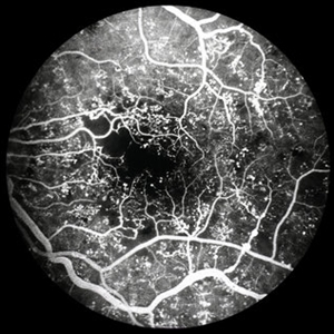
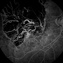
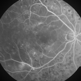
This is a fluorescein angiogram image capturing a beautiful example of different stages of venous beading in diabetic retinopathy all in one frame. This patient also has various microangiopathic findings including microaneurysms, venous loops and capillary dropout. This patient is a 41 y/o male with a history of type 1 diabetes, presenting for his first eye exam in years.
Imaging device: Optos FA
Condition/keywords: capillary dropouts, nonproliferative diabetic retinopathy, proliferative diabetic retinopathy (PDR), retinal ischemia, venous beading
-
 Diabetic Macular Edema, Proliferative Diabetic Retinopathy, Neovascularization Elsewhere, DME, PDR, NVE
Apr 1 2013 by James B. Soque, CRA, OCT-C, COA, FOPS
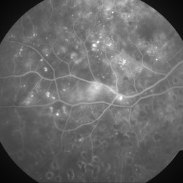
39-year-old white female and long standing diabetis, c/o new peripheral symptoms of left eye. FA OS reveals diabetic macular edema, microaneurysms, and neovasculaization elsewhere. Fluorescein Angogram, Early Phase, 50 Deg, 2x Mag.
Photographer: James B Soque, CRA, COA
Imaging device: Topcon TRC 50DX with MERGE software, OIS 10.6.45
Condition/keywords: diabetic macular edema, neovascularization (NV), proliferative diabetic retinopathy (PDR)
-
 Diabetic Retinopathy
Jun 4 2025 by Paulina Araujo
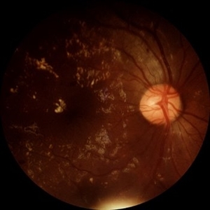
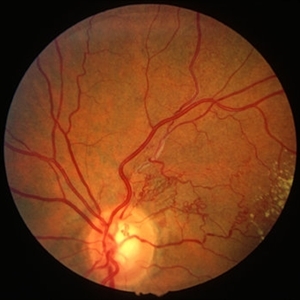
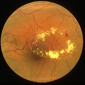
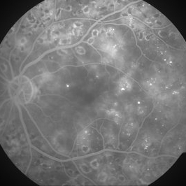
The 55-degree central fundus photograph of the right eye demonstrates numerous hard exudates, dot intraretinal hemorrhages, and microaneurysms.
Photographer: Paulina D.Araujo Martínez, Asociación para Evitar la Ceguera en México I.A.P., Hospital Dr Luis Sánchez Bulnes.
Condition/keywords: diabetic retinopathy
-
 Macular Pucker
Jan 7 2020 by RAFAEL REIS PEREIRA, MD
A clinical grading system was proposed by Gass in 1987 describe the different stages of the epiretinal membrane. Grade 2 Macular pucker consists of a thick fibroglial membrane that contracts and produces obscuration of underlying vessels and marked full-thickness retinal distortion. Sometimes associated with cotton-wool spots, exudates, blot hemorrhages, microaneurysms, and cystoid macular edema.
Photographer: Rafael Reis, Retina Clinic - Brazil
Condition/keywords: macular pucker
-
 NPDR
NPDR
Mar 29 2013 by Henry J. Kaplan, MD
Multiple microaneurysms visible as small round dot lesions.
Condition/keywords: nonproliferative diabetic retinopathy
-
 Proliferative Diabetic Retinopathy
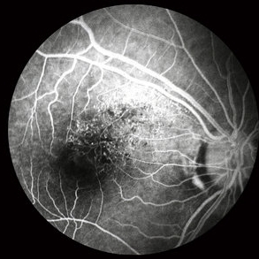
Oct 15 2012 by Susanna S. Park, MD, PhD
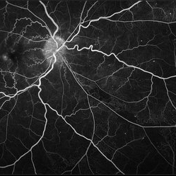
Fluorescein angiogram of the left eye of a 65 year old woman with diabetes mellitus showing nasal peripheral retinal capillary dropout and neovascularization of the disc. Scattered retinal microaneurysms are also noted
Photographer: Ellen Redenbo, University of California Davis Eye Center
Imaging device: Optos
Condition/keywords: proliferative diabetic retinopathy (PDR)
-
 Proliferative Diabetic Retinopathy
Aug 23 2012 by Gerardo Garcia-Aguirre, MD
Fluorescein angiogram of a left eye of a 45-year-old patient with proliferative diabetic retinopathy. Small hyperfluorescent dots are observed (microaneurysms), as well as blockage from a subhyaloid hemorrhage.
Photographer: Noemí Hernández, Asociación para Evitar la Ceguera en México
Condition/keywords: microaneurysms, subhyaloid hemorrhage
-
 Tractional Retinal Detachment
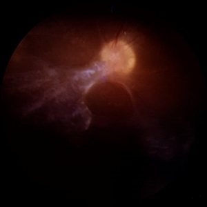
Jun 4 2025 by Paulina Araujo
The 55-degree central fundus photograph of the right eye reveals a thickened and opacified hyaloid exerting traction on the optic disc and posterior pole of the retina, along with hard exudates and microaneurysms consistent with advanced proliferative diabetic retinopathy.
Photographer: Paulina D.Araujo Martínez, Asociación para Evitar la Ceguera en México I.A.P., Hospital Dr Luis Sánchez Bulnes.
Condition/keywords: tractional retinal detachment
-
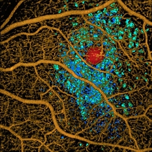 Volume Rendering Structural and Angiographic Optical Coherence Tomography Angiography Image of a Retinal Capillary Microaneurysm, A Newly Described Entity.
May 21 2019 by Richard F. Spaide, MD
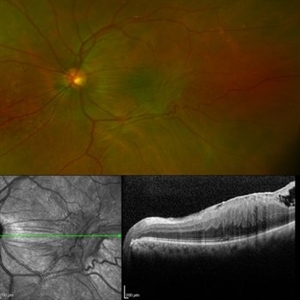
This is a newly described entity in which patients develop solitary aneurysms that are much larger than typical microaneurysms and they are supplied by capillaries. The aneurysm is shown in red. The associated macular edema produced cystoid spaces in Henle’s fiber layer, rendered as teal and in the inner nuclear layer as blue.
Photographer: Richard F. Spaide, MD
Condition/keywords: aneurysm, optical coherence tomography (OCT), volume rendering
-
 Proliferative Diabetic Retinopathy - Neovascularization on the Disc
Aug 23 2012 by Gerardo Garcia-Aguirre, MD
Fluorescein angiogram, early phase, showing microaneurysms, wide areas of capillary nonperfusion, and leakage secondary to neovascularization on the disc.
Photographer: Noemí Hernández, Asociación para Evitar la Ceguera en México
Condition/keywords: microaneurysms, neovascularization of the disc (NVD)
-
 BRVO FA, Early Phase
Oct 1 2012 by Jeffrey G. Gross, MD, FASRS
BRVO-FA early phase.
Condition/keywords: branch retinal vein occlusion (BRVO), capillary nonperfusion, early phase, microaneurysms
-
 PDR with Foveal Ischemia and FAZ Enlargement
Oct 8 2012 by Jeffrey G. Gross, MD, FASRS
PDR with foveal ischemia, and FAZ enlargement, multiple microaneurysms, FA, early phase.
Condition/keywords: early phase, enlarged foveal avascular zone, foveal ischemia, microaneurysms
-
 Proliferative Diabetic Retinopathy
Aug 23 2012 by Gerardo Garcia-Aguirre, MD
Fluorescein angiogram of a left eye of a 45 year-old patient with proliferative diabetic retinopathy. Small hyperfluorescent dots are observed (microaneurysms), as well as blockage from a subhyaloid hemorrhage. In the inferonasal area two areas of leakage secondary to neovascularization are observed.
Photographer: Noemí Hernández, Asociación para Evitar la Ceguera en México
Condition/keywords: microaneurysms, neovascularization (NV), subhyaloid hemorrhage
-
---thumb.jpg/image-square;max$300,300.ImageHandler) Binder3 P12 Slide82
Binder3 P12 Slide82
Feb 15 2013 by From the Collections of Thomas M. Aaberg, MD and Thomas M. Aaberg Jr., MD
Color fundus photograph showing peripheral retinal nonperfusion, retinal neovascularization elsewhere (NVE), venous beading and dilatation, retinal microaneurysms, and intraretinal hemorrhage.
Condition/keywords: peripheral retinal nonperfusion, proliferative retinopathy, retinal neovascularization
-
 Branch Retinal Vein Occlusion
Aug 22 2024 by Virginia Gebhart
Fluorescein angiogram of branch retinal vein occlusion in 75 year old female. Scattered microaneurysms with late CME and persistent SRF. Pt will consider laser treatment but is hesitant for injections at this time due to possible side effects.
Photographer: Virginia Gebhart
Imaging device: Optos California
Condition/keywords: branch retinal vein occlusion (BRVO), BRVO, cystoid macular edema (CME), FA, FA late phase, fluorescein angiogram (FA), macular edema, microaneurysms, retinal microaneurysms
-
 BRVO - longstanding
Jan 11 2013 by Alex P. Hunyor, MD
Longstanding branch retinal vein obstruction with collaterals, telangiectasia, microaneurysms and exudates - color image 1.
Condition/keywords: branch retinal vein occlusion (BRVO), collaterals
-
 BRVO - longstanding
Jan 11 2013 by Alex P. Hunyor, MD
Longstanding branch retinal vein obstruction with collaterals, telangiectasia, microaneurysms and exudates - color image 2.
Condition/keywords: branch retinal vein occlusion (BRVO)
-
 BRVO, FA, Hemorrhage, Diabetic
Mar 13 2014 by James B. Soque, CRA, OCT-C, COA, FOPS
51-year-old white male, diabetes, and with BRVO left eye, early phase 36 seconds. Flame heme from ON, showing microaneurysims, and fine capillary detail of this FA.
Photographer: James B Soque, CRA COA
Imaging device: Topcon TRC 50DX with MERGE software
Condition/keywords: branch retinal vein occlusion (BRVO), diabetes, FA early phase, microaneurysms
-
 Choroidal Melanoma - Stable, Fluorescein Angiogram, Early Phase
Mar 13 2019 by James B. Soque, CRA, OCT-C, COA, FOPS
Early FA, right eye, with choroidal melanoma-stable, and a few tiny microaneurysms showing leakage in re-circulation phase.
Photographer: James Soque, CRA, OCT-C, FOPS
Imaging device: Topcon TRC-50DX with MERGE Eye Station software
Condition/keywords: FA early phase, fluorescein angiogram (FA), MERGE, microaneurysms
-
 Chronic CRVO
Dec 12 2024 by Korey Starkey
Fluorescein Angiography of a 62 year-old man with chronic central retinal vein occlusion. Vision is 20/200.
Photographer: Korey Starkey
Imaging device: Optos
Condition/keywords: capillary nonperfusion, central retinal vein occlusion (CRVO), FLUORESCEIN ANGIOGRAPHY, ischemia, microaneurysms, Optos
-
 Diabetic Macular Edema - Leaking Microaneurysms
Oct 3 2013 by Gerardo Garcia-Aguirre, MD
Diabetic macular edema - leaking microaneurysms.
Condition/keywords: diabetic macular edema, diabetic retinopathy circinate
-
 Diabetic Macular Edema - Leaking Microaneurysms
Oct 3 2013 by Gerardo Garcia-Aguirre, MD
Diabetic macular edema - leaking microaneurysms.
Condition/keywords: diabetic macular edema
-
 Diabetic Macular Edema - Leaking Microaneurysms
Oct 3 2013 by Gerardo Garcia-Aguirre, MD
Diabetic macular edema - leaking microaneurysms.
Condition/keywords: diabetic macular edema
-
 Diabetic Macular Edema - Leaking Microaneurysms
Oct 3 2013 by Gerardo Garcia-Aguirre, MD
Diabetic macular edema - leaking microaneurysms.
Condition/keywords: diabetic macular edema
-
 Diabetic Retinopathy
Nov 20 2024 by Korey Starkey
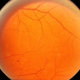
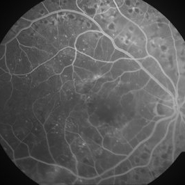
64 year old female being monitored for moderate-severe diabetic retinopathy.
Photographer: Korey Starkey
Condition/keywords: capillary nonperfusion, FA, FLUORESCEIN ANGIOGRAPHY, microaneurysms, nonproliferative diabetic retinopathy, Optos, OPTOS CALIFORNIA, tortuous vessels
 Loading…