Search results (73 results)
-
 Branch Retinal Vein Occlusion
Aug 22 2024 by Virginia Gebhart
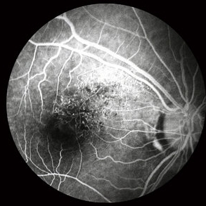
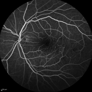
Fluorescein angiogram of branch retinal vein occlusion in 75 year old female. Scattered microaneurysms with late CME and persistent SRF. Pt will consider laser treatment but is hesitant for injections at this time due to possible side effects.
Photographer: Virginia Gebhart
Imaging device: Optos California
Condition/keywords: branch retinal vein occlusion (BRVO), BRVO, cystoid macular edema (CME), FA, FA late phase, fluorescein angiogram (FA), macular edema, microaneurysms, retinal microaneurysms
-
 BRVO FA, Early Phase
Oct 1 2012 by Jeffrey G. Gross, MD, FASRS
BRVO-FA early phase.
Condition/keywords: branch retinal vein occlusion (BRVO), capillary nonperfusion, early phase, microaneurysms
-
 BRVO, FA, Hemorrhage, Diabetic
Mar 13 2014 by James B. Soque, CRA, OCT-C, COA, FOPS
51-year-old white male, diabetes, and with BRVO left eye, early phase 36 seconds. Flame heme from ON, showing microaneurysims, and fine capillary detail of this FA.
Photographer: James B Soque, CRA COA
Imaging device: Topcon TRC 50DX with MERGE software
Condition/keywords: branch retinal vein occlusion (BRVO), diabetes, FA early phase, microaneurysms
-
 Choroidal Melanoma - Stable, Fluorescein Angiogram, Early Phase
Mar 13 2019 by James B. Soque, CRA, OCT-C, COA, FOPS
Early FA, right eye, with choroidal melanoma-stable, and a few tiny microaneurysms showing leakage in re-circulation phase.
Photographer: James Soque, CRA, OCT-C, FOPS
Imaging device: Topcon TRC-50DX with MERGE Eye Station software
Condition/keywords: FA early phase, fluorescein angiogram (FA), MERGE, microaneurysms
-
 Chronic CRVO
Dec 12 2024 by Korey Starkey
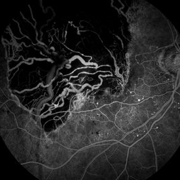
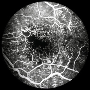
Fluorescein Angiography of a 62 year-old man with chronic central retinal vein occlusion. Vision is 20/200.
Photographer: Korey Starkey
Imaging device: Optos
Condition/keywords: capillary nonperfusion, central retinal vein occlusion (CRVO), FLUORESCEIN ANGIOGRAPHY, ischemia, microaneurysms, Optos
-
 Diabetic Retinopathy
Nov 20 2024 by Korey Starkey
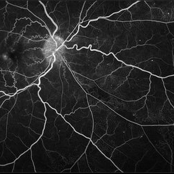
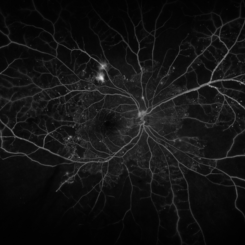
64 year old female being monitored for moderate-severe diabetic retinopathy.
Photographer: Korey Starkey
Condition/keywords: capillary nonperfusion, FA, FLUORESCEIN ANGIOGRAPHY, microaneurysms, nonproliferative diabetic retinopathy, Optos, OPTOS CALIFORNIA, tortuous vessels
-
 Fluorescein Angiogram - Tortuous Vessels of DME Right Eye
Dec 10 2015 by James B. Soque, CRA, OCT-C, COA, FOPS
Early fluorescein angiogram of diabetic macular edema and tortuous vessels in the superior macula of the right eye.
Photographer: James B Soque, CRA, COA
Imaging device: Top[con TRC-50 DX with MERGE Winstation V 11.2.0
Condition/keywords: diabetes, diabetic macular edema, microaneurysms, microangiopathy, tortuous vessels
-
 Idiophatic retinal vasculitis
Jul 9 2023 by Luiz A Zago, PhD
Mid phase angiography of a 45 woman with a idiophatic vasculitis. She is been followed in the last 10 years. No sistemic association was found beside face rosacea, This case was first described in Diagnostic and Therapeutic Challenges - Retina Journal
Photographer: Luiz Zago, PhD.
Imaging device: Topcon 50IX
Condition/keywords: anomalous foveal avascular zone, Choroidal Folds, intraretinal microvascular abnormalities, microaneurysms, optic neuritis, Vaculitis
-
 Neovascularization of the Disc
Jun 3 2025 by Scott D Walter, MD, MSc, FASRS
Near-infrared (NIR) en face OCT image showing neovascularization of the disc (NVD) in a patient with type II diabetes mellitus, complicated by proliferative diabetic retinopathy (PDR).
Imaging device: Heidelberg Spectralis
Condition/keywords: Diabetes, Heidelburg Spectralis, microaneurysms, Neovascularisation at the Disc (NVD), NEOVASCULARISATION OF DISC, OCT EN FACE, proliferative diabetic retinopathy (PDR)
-
 Old Macular BRVO
Oct 10 2015 by Hamid Ahmadieh, MD
Mid -venous phase FA of the left eye of a 58-year-old woman with the history of macular BRVO. Note microaneurysms, collaterals , tortuosity of venules and an enlarged irregular FAZ compatible with macular ischemia.
Photographer: Nayereh Hadipour, Negah Eye Center, Tehran, Iran
Condition/keywords: collaterals, macular branch retinal vein occlusion (BRVO), macular ischemia, microaneurysms
-
 PDR with Foveal Ischemia and FAZ Enlargement
Oct 8 2012 by Jeffrey G. Gross, MD, FASRS
PDR with foveal ischemia, and FAZ enlargement, multiple microaneurysms, FA, early phase.
Condition/keywords: early phase, enlarged foveal avascular zone, foveal ischemia, microaneurysms
-
 PDR with Ischemia
Jul 7 2020 by Stephanie Burke
Early frame of a 45-year-old male with Type II diabetes.
Photographer: Stephanie Burke, CRA, OCT-C
Condition/keywords: FA early phase, ischemia, microaneurysms, neovascularization (NV), proliferative diabetic retinopathy (PDR), ultra-wide field imaging, venous beading
-
 Proliferative Diabetic Retinopathy
Mar 3 2017 by Nichole Lewis
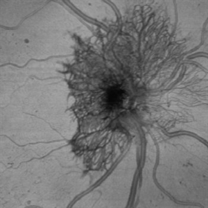
61-year-old female with proliferative diabetic retinopathy, neovascularization, microaneurysms, dot blot hemorrhages and capillary nonperfusion.
Photographer: Nichole Lewis
Condition/keywords: microaneurysms, neovascularization elsewhere (NVE), proliferative diabetic retinopathy (PDR)
-
 Proliferative Diabetic Retinopathy
Apr 29 2020 by Stephanie Burke
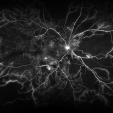
Ultra-wide FA image of a 58-year-old man with DM.
Photographer: Stephanie Burke, CRA, OCT-C
Condition/keywords: ischemia, leakage, microaneurysms, neovascularization (NV), proliferative diabetic retinopathy (PDR), ultra-wide field imaging
-
 Proliferative Diabetic Retinopathy
Aug 23 2012 by Gerardo Garcia-Aguirre, MD
Fluorescein angiogram of a left eye of a 45-year-old patient with proliferative diabetic retinopathy. Small hyperfluorescent dots are observed (microaneurysms), as well as blockage from a subhyaloid hemorrhage.
Photographer: Noemí Hernández, Asociación para Evitar la Ceguera en México
Condition/keywords: microaneurysms, subhyaloid hemorrhage
-
 Proliferative Diabetic Retinopathy
Aug 23 2012 by Gerardo Garcia-Aguirre, MD
Fluorescein angiogram of a left eye of a 45 year-old patient with proliferative diabetic retinopathy. Small hyperfluorescent dots are observed (microaneurysms), as well as blockage from a subhyaloid hemorrhage. In the inferonasal area two areas of leakage secondary to neovascularization are observed.
Photographer: Noemí Hernández, Asociación para Evitar la Ceguera en México
Condition/keywords: microaneurysms, neovascularization (NV), subhyaloid hemorrhage
-
 Proliferative Diabetic Retinopathy - Neovascularization on the Disc
Aug 23 2012 by Gerardo Garcia-Aguirre, MD
Fluorescein angiogram, early phase, showing microaneurysms, wide areas of capillary nonperfusion, and leakage secondary to neovascularization on the disc.
Photographer: Noemí Hernández, Asociación para Evitar la Ceguera en México
Condition/keywords: microaneurysms, neovascularization of the disc (NVD)
-
 Proliferative Diabetic Retinopathy - Neovascularization on the Disc
Aug 23 2012 by Gerardo Garcia-Aguirre, MD
Fluorescein angiogram, late phase, showing microaneurysms, wide areas of capillary non-perfusion, and leakage secondary to neovascularization on the disc.
Photographer: Noemí Hernández, Asociación para Evitar la Ceguera en México
Condition/keywords: microaneurysms, neovascularization of the disc (NVD)
-
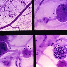 Slide 9-15
Feb 26 2019 by Lancaster Course in Ophthalmology
Trypsin digest retinal preparation showing diabetic microaneurysms. Some microaneurysms are relatively acellular (upper right and lower left), and others are filled with neutrophils (lower right).
Condition/keywords: microaneurysms, neutrophils, trypsin digestion
-
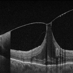 Vitreomacular Adhesion Showcasing a Microaneurysm and a Subhyaloid Hemorrhage
Jan 3 2025 by Drew Mitchell
A vertical OCT 1 line raster scan positioned slightly inferomacula to document the subhyaloid hemorrhage. Hyper reflective Oval indicating Microaneurysm.
Photographer: Drew Mitchell, OCT-C
Imaging device: Zeiss Cirrus 5000
Condition/keywords: diabetic macular edema, microaneurysms, retinal microaneurysms, subhyaloid hemorrhage, subretinal fluid, vitreomacular adhesion, vitreomacular traction (VMT)
-
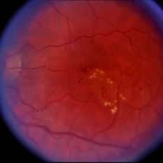
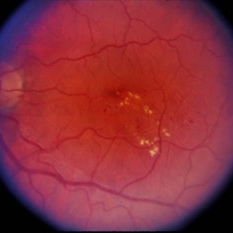 Perifoveal Telangiectasia
Jan 5 2015 by H. Michael Lambert, MD
Microaneurysms in the temporal foveal area with lipid; Left eye.
Condition/keywords: perifoveal telangiectasia
-
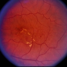
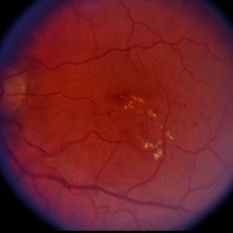 Perifoveal Telangiectasia
Jan 5 2015 by H. Michael Lambert, MD
Microaneurysms in the temporal foveal area with lipid; Left eye.
Condition/keywords: perifoveal telangiectasia
-
 Perifoveal Telangiectasia
Jan 5 2015 by H. Michael Lambert, MD
Microaneurysms in the temporal foveal area with lipid; Left eye.
Condition/keywords: perifoveal telangiectasia
-
 Perifoveal Telangiectasia
Jan 5 2015 by H. Michael Lambert, MD
Microaneurysms in the temporal foveal area with lipid; Left eye.
Condition/keywords: perifoveal telangiectasia
-
---thumb.jpg/image-square;max$300,300.ImageHandler) Binder3 P12 Slide82
Binder3 P12 Slide82
Feb 15 2013 by From the Collections of Thomas M. Aaberg, MD and Thomas M. Aaberg Jr., MD
Color fundus photograph showing peripheral retinal nonperfusion, retinal neovascularization elsewhere (NVE), venous beading and dilatation, retinal microaneurysms, and intraretinal hemorrhage.
Condition/keywords: peripheral retinal nonperfusion, proliferative retinopathy, retinal neovascularization
 Loading…