Search results (264 results)
-
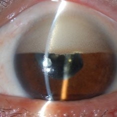
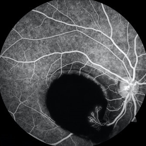 Sub-ILM Hemorrhage with Neovessels
Apr 30 2020 by Saurabh Deshmukh, MBBS, DNB, FVRS, MNAMS
Late arteriovenous phase FA showing a large sub-internal limiting membrane hemorrhage with overlying neovessels. This hypertensive patient presented with a visual acuity of counting fingers at 2 meters. The patient was advised intravitreal anti-VEGF injection, Nd: YAG Membranotomy, and systemic control of hypertension.
Photographer: Saurabh Deshmukh, Sri Sankaradeva Nethralaya, Guwahati, India
Imaging device: Topcon TRC-50 DX
Condition/keywords: hypertensive retinopathy, neovascularization elsewhere (NVE), subILM hemorrhage
-
 Venous Loop & Venous Beading
May 31 2014 by Hamid Ahmadieh, MD
Color fundus photograph of the left eye of a diabetic patient with NVD, NVE, venous loop and venous beading.
Photographer: Elham Salehi, Negah Eye Center, Tehran
Condition/keywords: color fundus photograph, neovascularization elsewhere (NVE), neovascularization of the disc (NVD), proliferative diabetic retinopathy (PDR), venous beading, venous loop
-
 Scleral Indentation In A Normal Eye
Nov 9 2012 by Norman Byer
This shows the appearance of scleral indentation in a normal eye. Note the convex shadow which marks the posterior border of the indented area. It is caused in part by a small angle which separates the viewing axis from the illuminating axis thus allowing the observer to see slightly into the shadow beyond the illuminated crest of the indentation. It is also caused in part by viewing the pigment epithelial layer in a tangential manner. This shadow is of great diagnostic usefulness since it becomes a dark background against which many tiny retinal abnormalities can be seen beautifully by contrast. Two other particular advantages of scleral indentation will be demonstrated in the following photographs: First, the ability to see the extreme anterior part of the retina to the ora serrata and beyond, and second, the ability to examine any abnormality in multiple profiles depending on slight movements of the scleral depressor in various directions.
Condition/keywords: extreme anterior retina, posterior border, scleral indentation, shadow, tangential view of pigment epithelial layer
-
 Giant Papillary Conjunctivitis
Dec 13 2013 by Jason S. Calhoun
Patient wears soft contact lenses complained of irritation when the SCL would move. Inverted eyelid in both eyes and there was papillary +2 underneath the eyelid.
Photographer: Jason S. Calhoun, Ophthalmic Photographer, Department of Ophthalmology, Mayo Clinic Jacksonville
Imaging device: TOPCON D-90 SL NIKON CAMERA
Condition/keywords: giant papillary conjunctivitis
-
 Diabetic Macular Edema, Proliferative Diabetic Retinopathy, Neovascularization Elsewhere, DME, PDR, NVE
Apr 1 2013 by James B. Soque, CRA, OCT-C, COA, FOPS
39-year-old white female and long standing diabetis, c/o new peripheral symptoms of left eye. FA OS reveals diabetic macular edema, microaneurysms, and neovasculaization elsewhere. Fluorescein Angogram, Early Phase, 50 Deg, 2x Mag.
Photographer: James B Soque, CRA, COA
Imaging device: Topcon TRC 50DX with MERGE software, OIS 10.6.45
Condition/keywords: diabetic macular edema, neovascularization (NV), proliferative diabetic retinopathy (PDR)
-
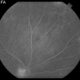
 Sea Fan Neovascularisation
Apr 27 2015 by Neha Goel, MS DNB FRCS (Glasg)
Fluorescein angiography of the left eye of a 40-year-old male.
Photographer: Neha Goel
Imaging device: Zeiss visucam
Condition/keywords: Eales disease, neovascularization elsewhere (NVE), vasculitis
-
 Giant Papillary Conjunctivitis
Dec 13 2013 by Jason S. Calhoun
Patient wears soft contact lenses complained of irritation when the SCL would move. Inverted eyelid in both eyes and there was papillary +2 underneath the eyelid.
Photographer: Jason S. Calhoun, Ophthalmic Photographer, Department of Ophthalmology, Mayo Clinic Jacksonville
Imaging device: TOPCON D-90 SL NIKON CAMERA
Condition/keywords: giant papillary conjunctivitis
-
 Choroidal Detachment
Mar 29 2013 by Henry J. Kaplan, MD
One quadrant choroidal detachment as brownish convex lesion.
Condition/keywords: choroidal detachment
-
 Venous Loop & Venous Beading
May 31 2014 by Hamid Ahmadieh, MD
Color fundus photograph of the left eye of a diabetic patient with NVD, NVE, venous loop and venous beading.
Photographer: Elham Salehi, Negah Eye Center, Tehran
Condition/keywords: color fundus photograph, neovascularization elsewhere (NVE), neovascularization of the disc (NVD), proliferative diabetic retinopathy (PDR), venous beading, venous loop
-
---thumb.JPG/image-square;max$300,300.ImageHandler) Proliferative Diabetic Retinopathy With Neovascularization - Red-Free
Proliferative Diabetic Retinopathy With Neovascularization - Red-Free
Jan 31 2014 by Roy Schwartz, MD
Red-free image of a 57-year-old woman with proliferative diabetic retinopathy, including NVD and NVE
Photographer: Galit Yair Pur
Condition/keywords: neovascularization (NV), proliferative diabetic retinopathy (PDR), red-free
-
 Inverted Hypopyon - Silicon Oil Complication
Feb 12 2015 by H. Michael Lambert, MD
Silicon oil emulsification, inverted hypopyon.
Condition/keywords: hypopyon, silicone oil
-
 Elevated Lesion
Nov 9 2012 by Norman Byer
This photograph and the next are two views of a very interesting elevated lesion in a 45-year-old man. This photograph shows the immense value of closely scrutinizing the profile of the indented area. Note that in the middle of the slide there is a sudden break in the continuity of the dark convex shadow that lies just behind the crest of the scleral indentation. If the elevated tissue is "filmy" or "wispy" or filamentous as in this case, it raises a strong suspicion that a retinal break is present just behind it.
Condition/keywords: elevated retinal lesion, elevated tissue, retinal break, scleral indentation
-
 Venous Beading & NVE
Mar 29 2013 by Henry J. Kaplan, MD
Typical venous beading and NVE in a diabetic patient with PDR.
Condition/keywords: neovascularization (NV), venous beading
-
 Advanced Active PDR
Mar 29 2013 by Henry J. Kaplan, MD
Extensive NVD-FPD and NVE-FPE in a diabetic patient.
Condition/keywords: foveal photoreceptor defect, FPE, neovascularization (NV), neovascularization of the disc (NVD)
-
 Retinoschisis Detachment
Nov 9 2012 by Norman Byer
Combined retinoschisis detachment, so-called schisis detachment, in a 47-year-old woman. The large outer layer hole in the center has a posterior yellow border which represents the position of the outer layer. Please observe superior to the hole the dark convexity of the scleral indentation. Just below the hole at the middle of the slide and going to the left the yellow zone comes to lie right against the inner layer and a fluid filled cavity lies deep to the outer layer. At this point, therefore, there is a true neurosensory detachment of the retina. On the right side of the hole, the yellow line slants up and to the right and lies close to the pigment epithelium. On the right side of the photograph, the original schisis cavity can be seen separating the yellow line of the outer layer above from the inner retinal layer below. The mechanism of this detachment is that some of the fluid from the schisis cavity passes through the outer layer hole and detaches the outer layer. This lesion has not been treated and has remained exactly the same for 13 years. A similar symmetrical "schisis-detachment" is present in the fellow eye.
Condition/keywords: neurosensory detachment of retina, outer layer hole, pigment epithelium, retinoschisis, schisis detachment, scleral indentation
-
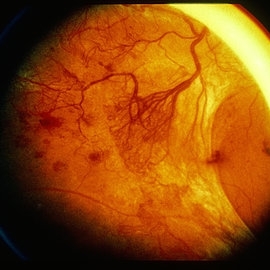
 Marked Retinal Ischemia in Patient with Mixed Connective Tissue Disease
Feb 26 2013 by Sharon Fekrat, MD FACS FASRS
Fluorescein angiogram of the right eye of a 27-year-old female with mixed connective tissue disease and marked retinal ischemia. Panretinal laser photocoagulation (PRP) has been performed for neovascularization elsewhere (NVE).
Condition/keywords: mixed connective tissue disease, retinal ischemia
-
 Inverted Hypopyon - Silicon Oil Complication
Feb 12 2015 by H. Michael Lambert, MD
Silicon oil emulsification, inverted hypopyon.
Condition/keywords: hypopyon, silicone oil
-
 Retinoschisis
Nov 9 2012 by Norman Byer
This 53-year-old man has retinoschisis involving the upper temporal quadrant but with no visible yellow dots or white lines to make it obvious. However, with scleral indentation you can see a large convex area showing the so-called white with pressure phenomenon. This area corresponds exactly to the area being indented and therefore must arise either from the outer layer of the retina or from some structure deep to it. White with pressure is an interesting optical phenomenon of uncertain origin but of no definite diagnostic or prognostic significance.
Condition/keywords: retinoschisis, scleral indentation, white with pressure
-
 Inverse Hypopyon
Mar 4 2018 by Yoshihiro Yonekawa, MD, FASRS
Slit lamp photograph of a 40-year-old man with previous retinal detachment surgery with silicone oil tamponade, presenting with an inverse hypopyon from emulsified silicone oil.
Photographer: Steven A Bennett, COA, CRA
Imaging device: Nikon D200 / Topcon Slit lamp
Condition/keywords: hypopyon, silicone oil
-
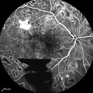
 PDR NVD NVE
Jul 21 2014 by Susanna S. Park, MD, PhD
Mid-transit view fluorecein angiogram of the right eye of a 59-year-old diabetic woman with minimal peripheral fundus changes suggestive of diabetic retinopathy showing diffuse leakage of the disc and focal leakage in the peripheral retina from neovascularization. Peripheral retinal ischemia and leaking retinal microaneurysms are also seen.
Photographer: Karishma Chandra, University of California Davis Eye Center
Condition/keywords: fluorescein leakage, neovascularization of the disc (NVD), proliferative diabetic retinopathy (PDR)
-
 Advanced Active PDR
Mar 29 2013 by Henry J. Kaplan, MD
Large active NVEs with fibrous proliferations in diabetes.
Condition/keywords: fibrous proliferation, neovascularization (NV)
-
 Giant Retinal Tear
Mar 29 2014 by Min Kim, MD, PhD, MBA, FASRS
Wide field fundus photograph of a 25 year-old male shows giant retinal tear with inverted retinal flap.
Photographer: Young Duk Bae, Yonsei University, Gangnam Severance Hospital
Imaging device: Optomap
Condition/keywords: giant retinal tear
-
 Proliferative Diabetic Retinopathy With Neovascularization - FA
Jan 31 2014 by Roy Schwartz, MD
Fluorescein angiogram of a 57-year-old woman with proliferative diabetic retinopathy, including NVD and NVE
Photographer: Galit Yair Pur
Condition/keywords: neovascularization (NV), proliferative diabetic retinopathy (PDR)
-
 Proliferative Diabetic Retinopathy With Neovascularization - FA
Jan 31 2014 by Roy Schwartz, MD
Fluorescein angiogram of a 57-year-old woman with proliferative diabetic retinopathy, including NVD and NVE.
Photographer: Galit Yair Pur
Condition/keywords: neovascularization (NV), proliferative diabetic retinopathy (PDR)
-
 Proliferative Diabetic Retinopathy
Sep 15 2012 by Hamid Ahmadieh, MD
FA image of a 30-year-old woman with the history of scatter laser photocoagulation, NVE and a preretinal hemorrhage due to active PDR .
Photographer: Hamid Ahmadieh, MD, Ophthalmic Research Center, Labbafinejad Medical Center, Shahid Beheshti University of Medical Sciences
Imaging device: Heidelberg HRA
Condition/keywords: preretinal hemorrhage
 Loading…