Search results (264 results)
-
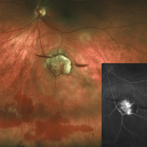
 Bilateral Proliferative Diabetic Retinopathy OU
Feb 21 2025 by Drew Mitchell
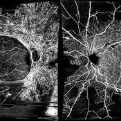
OCT-Angiography 8x8 Montage OU. PDR with active NVE OD. 37 year old male with no visual complaints. Vision is 20/20 in both eyes.
Photographer: Drew Mitchell OCT-C
Imaging device: Zeiss Cirrus 5000
Condition/keywords: CIRRUS 5000 ANGIOPLEX, Diabetes, NVE, OCT Angiography, proliferative diabetic retinopathy (PDR)
-
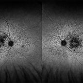
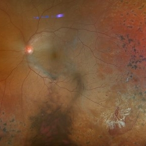
 Branches Starved of Flow, Yet Nature Strives to Grow
Apr 1 2025 by rohan jain
Tufts of NVE's in a case of Branch Retinal Vein Occlusion
Photographer: Dr. ROHAN JAIN
Condition/keywords: branch retinal vein occlusion (BRVO), capillary nonperfusion, non-perfused branch retinal vein occlusion (BRVO), non-perfusion, NVE, OCT Angiography, ST BRVO
-
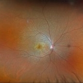 Multifocal Pattern Dystrophy
Feb 5 2025 by Kimberly Wakester
Optomap RGB and AF photograph of an 37-year-old woman with multifocal pattern dystrophy in both eyes. Previously believed to be Stargardts, but genetic testing returned positive for PRPH2 mutation. Likely Multifocal Pattern Dystrophy given phenotypical appearance of SGD. There is stable NVE in the left eye. Will continue to monitor both eyes and consider treatment with PRP laser if needed for NVE in the left eye.
Photographer: Kimberly Wakester, COA
Imaging device: Optos California
Condition/keywords: multifocal pattern dystrophy, NVE, PRPH2 Positive
-
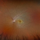 Multifocal Pattern Dystrophy
Feb 5 2025 by Kimberly Wakester
Optomap RGB and AF photograph of an 37-year-old woman with multifocal pattern dystrophy in both eyes. Previously believed to be Stargardts, but genetic testing returned positive for PRPH2 mutation. Likely Multifocal Pattern Dystrophy given phenotypical appearance of SGD. There is stable NVE in the left eye. Will continue to monitor both eyes and consider treatment with PRP laser if needed for NVE in the left eye.
Photographer: Kimberly Wakester, COA
Imaging device: Optos California
Condition/keywords: multifocal pattern dystrophy, NVE, PRPH2 Positive
-
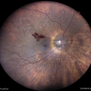 Neovascularization Elsewhere in a Case of Proliferative Diabetic Retinopathy
May 7 2024 by Akansha Sharma
Color fundus photograph of a 65 year old female with neovascularization elsewhere in a case of proliferative diabetic retinopathy.
Photographer: Dr. Akansha Sharma, Bharati Eye Hospital
Condition/keywords: NVE, PDR, proliferative diabetic retinopathy (PDR)
-
---thumb.jpg/image-square;max$300,300.ImageHandler) NVE
NVE
Feb 13 2013 by From the Collections of Thomas M. Aaberg, MD and Thomas M. Aaberg Jr., MD
Severe NV; ischemia, Pt GL
Condition/keywords: ischemia, neovascularization (NV)
-
 PDR
PDR
Mar 15 2024 by Virginia Gebhart
FA of 59 year old female with proliferative diabetic retinopathy.
Photographer: Virginia Gebhart
Imaging device: Optos California
Condition/keywords: Diabetic Retinopathy, neovascularization of the disc (NVD), NVE, proliferative diabetic retinopathy (PDR)
-
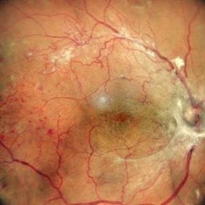 Proliferative Diabetic Retinopathy
May 24 2024 by Anjana Mirajkar, MS Ophthalmology
A central photo of a 65 year old female of right eye showing fibro vascular proliferation with neovascularization elsewhere in a case of proliferative diabetic retinopathy.
Photographer: Dr. Anjana Mirajkar -Retina Foundation, Ahmedabad
Imaging device: Mirante-Nidek
Condition/keywords: NVE, proliferative diabetic retinopathy (PDR), tractional retinal detachment
-
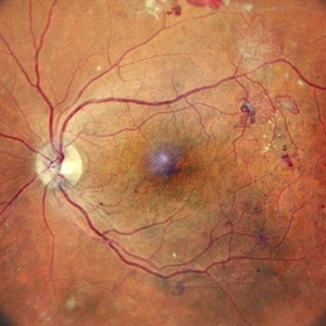 Proliferative Diabetic Retinopathy
May 24 2024 by Anjana Mirajkar, MS Ophthalmology
A central photo of a 60 year old male of left eye case of neovascularization elsewhere with dot and blot hemorrhages in a case of proliferative diabetic retinopathy.
Photographer: Dr. Anjana Mirajkar -Retina Foundation, Ahmedabad
Imaging device: Mirante-Nidek
Condition/keywords: NVE, pre-proliferative diabetic retinopathy
-
 Proliferative Diabetic Retinopathy- Red free
Dec 21 2023 by Vishal Agrawal, MD, FRCS,FACS,FASRS
20-year-old male patient having PDR with multiple NVE in all quadrants. Red free image accentuates the NVE as compared to color pic.
Photographer: Dr Ayushi
Imaging device: Clarus 700
Condition/keywords: NVE, PDR, RedFree
-
 Systemic Lupus Erythematosus (SLE) Vasculitis
Jan 29 2025 by Kimberly Wakester
Fundus photographs of an 13-year-old boy with Systemic Lupus Erythematosus (SLE) Vasculitis in both eyes s/p PRP laser. Patient is doing well s/p PRP Laser OU and with continued use of oral medications. Patient will be monitored with follow up exams to check for recurring vasculitis or recurring/worsening NVE/NVD. Patient is to continue ongoing management with Rheumatologist.
Photographer: Kimberly Wakester, COA
Imaging device: Optos California
Condition/keywords: NVD, NVE, occlusive vasculitis, pan-retinal photocoagulation (PRP), Systemic Lupus Erythematosus (SLE) Vasculitis
-
 NVE from BRVO
Feb 19 2015 by H. Michael Lambert, MD
Color photo of NVE after BRVO.
Condition/keywords: branch retinal vein occlusion (BRVO), neovascularization elsewhere (NVE)
-
 NVE from BRVO
Feb 19 2015 by H. Michael Lambert, MD
Color photo of NVE after BRVO.
Condition/keywords: branch retinal vein occlusion (BRVO), neovascularization elsewhere (NVE)
-
 NVE from BRVO
Feb 19 2015 by H. Michael Lambert, MD
Color photo of NVE after BRVO.
Condition/keywords: branch retinal vein occlusion (BRVO), neovascularization elsewhere (NVE)
-
 NVE in a Patient With Vasculitis
Nov 5 2018 by awaneesh m upadhyay, MBBS, DNB
FFA image of a 22-year-old male vasculitis patient with NVE.
Photographer: Hiteshwar Saikia
Condition/keywords: neovascularization elsewhere (NVE), tuberculosis, vasculitis
-
 NVE in an HIV Positive Case
Sep 26 2021 by Nivesh Gupta
FA Montage of a 22-year-old female with Neovascularization Elsewhere
Photographer: DR. NIVESH GUPTA, RETINA FOUNDATION, AHMEDABAD
Imaging device: NIDEK MIRANTE
Condition/keywords: HIV, neovascularization elsewhere (NVE)
-
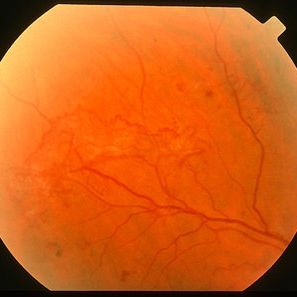 NVE in PDR - colour image
Jan 11 2013 by Alex P. Hunyor, MD
Extensive patch of NVE in proliferative diabetic retinopathy - color image.
Condition/keywords: neovascularization (NV)
-
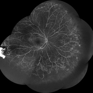
 NVE in PDR - FA
Sep 30 2013 by Alex P. Hunyor, MD
Extensive patch of NVE in proliferative diabetic retinopathy-fluorescein angiogram.
Condition/keywords: neovascularization (NV)
-
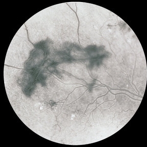
 NVE with Hemorrhages
Jan 20 2020 by Sarah Oelrich
NVE with hemorrhages
Photographer: Sarah Oelrich CRA COT OCT-C Southeastern Retina Associates
Condition/keywords: hemorrhage, neovascularization elsewhere (NVE)
-
 Active neovascularization in Proliferative Diabetic Retinopathy
Jan 10 2018 by Peter H. Tang, MD, PhD
Fluorescein angiography image from a 46-year-old woman with uncontrolled proliferative diabetic retinopathy shows extensive dye leakage from active neovascularization.
Imaging device: Optos California
Condition/keywords: diabetes, diabetic retinopathy, fluorescein leakage, neovascularization elsewhere (NVE), neovascularization of the disc (NVD), pan-retinal photocoagulation (PRP), proliferative diabetic retinopathy (PDR)
-
 Active Vasculitis with Proliferative Retinopathy
Jan 30 2021 by Raja Rami P Reddy, MD FRCS FASRS
25-year-old boy with unilateral recent onset visual loss. Fundus shows areas of active vasculitis nasally and large neovascular complexes temporally and on the disc and early fibrous membrane formation. Fellow eye fundus is normal. Further investigations suggested tubercular etiology
Photographer: Raja Rami Reddy P
Imaging device: fundus camera
Condition/keywords: proliferative retinopathy, tuberculosis, vasculitis
-
 Actively Bleeding NVE
Apr 1 2025 by Jordyn Beckman
47 year old woman presented with actively bleeding NVE temporally on exam with complaints of foggy vision and floaters.
Photographer: Jordyn Beckman, Retina Consultants of Carolina, P.A.
Imaging device: Optos California
Condition/keywords: active bleeding, Elevated retinal neovascularization, vitreous hemorrhage
-
 acute posterior multifocal placoid pigment epitheliopathy
Sep 23 2022 by Jaideep sharma
A 50-year old woman presented to us with unilateral progressive and painless visual blurring. She was diagnosed as a case of CSCR and started on topical dorzolamide with no improvement in VA. Her best-corrected visual acuity (BCVA) was RE 6/6 and LE 6/60 . Eye examination revealed vitritis (grade1) with optic disc hyperemia and multiple serous retinal detachments with choroidal striae in the left eye and a normal right eye. She is k/c/o diabetes. Her past ocular and drug histories were unremarkable. Retinal imaging revealed characteristic features of APMPPE in the left eye. All laboratory testing results were inconclusive. VA and OCT findings significantly improved following the treatment with LE posterior sub tenon’s triamcinolone (40 mg/ml). 1 month post injection VA of the left eye reached 6/6 with resolved serous retinal detachments in this eye. This case is unique as it was managed via PST injection rather than conventional steroid therapy
Photographer: jaideep sharma jaipur calgary eye hospital rajasthan india
Condition/keywords: acute posterior multifocal placoid pigment epitheliopathy (APMPPE), FFA
 Loading…