 Initializing download.
-
 By Norman Byer
From Dr. Norman E. Byer’s “The Peripheral Retina in Profile” - Uploaded on Nov 9, 2012.
- Last modified by Suber S. Huang, MD, MBA, FASRS on Feb 11, 2013.
- Reviewed by Chayal Patel
- Rating
- Appears in
- Miscellaneous
- Condition/keywords
- retinoschisis, schisis detachment, pigment epithelium, outer layer hole, scleral indentation, neurosensory detachment of retina
- Description
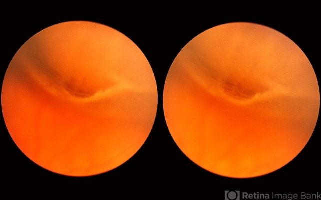
- Combined retinoschisis detachment, so-called schisis detachment, in a 47-year-old woman. The large outer layer hole in the center has a posterior yellow border which represents the position of the outer layer. Please observe superior to the hole the dark convexity of the scleral indentation. Just below the hole at the middle of the slide and going to the left the yellow zone comes to lie right against the inner layer and a fluid filled cavity lies deep to the outer layer. At this point, therefore, there is a true neurosensory detachment of the retina. On the right side of the hole, the yellow line slants up and to the right and lies close to the pigment epithelium. On the right side of the photograph, the original schisis cavity can be seen separating the yellow line of the outer layer above from the inner retinal layer below. The mechanism of this detachment is that some of the fluid from the schisis cavity passes through the outer layer hole and detaches the outer layer. This lesion has not been treated and has remained exactly the same for 13 years. A similar symmetrical "schisis-detachment" is present in the fellow eye.


")


---thumb.jpg/image-square;max$79,0.ImageHandler "Retinoschisis")


