Search results (264 results)
-
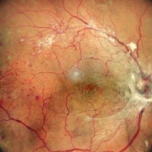
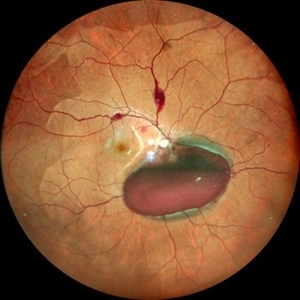 Valsalva Retinopathy
Dec 20 2021 by Unnati Vishwanath Shukla, M. S. ,DNB, FVRS FNERF, MNAMS,PhD Scholar(Retina)
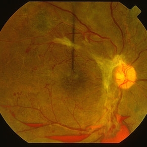
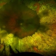
26-year-old male with Valsalva Retinopathy. History of severe cough for 3 days. All hematological investigations were within normal limits.
Photographer: Dr. Unnati Shukla, Consultant, Retina Foundation, Ahmedabad
Imaging device: Nidek Mirante
Condition/keywords: subhyaloid hemorrhage, subretinal hemorrhage, valsalva retinopathy
-
 Central Retinal Artery Occlusion With Cilioretinal Sparing
Apr 4 2018 by Soumya Venkatesh
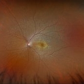
Fundus photograph of a 23-year-old gentleman presenting with sudden loss of vision 2 days prior to presentation. He underwent all relevant investigations and found to have APLA positive. He also had dengue serology positive. On follow up, his retinal edema reduced unmasking the underlying hemorrhages( flame shaped).
Photographer: Soumya Harapanahalli Venkatesh, JSS university, Karnataka, India
Condition/keywords: central retinal artery occlusion (CRAO), cherry red spot, cilioretinal sparing, retinal ischemia
-
Eye of the Hurricane
Apr 9 2025 by Gustavo Uriel Fonseca Aguirre
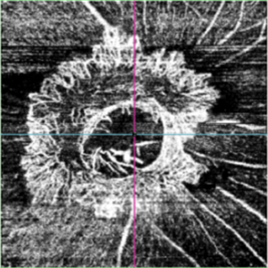
Ultrasound biomicroscopy of a post-operative eye (status post trabeculectomy and phacoemulsification) reveals a patent ostium on the right side, along with an intraocular lens in position. A hyphema is observed displaying small convection currents, creating a circular motion pattern due to the temperature gradient between the iris and cornea. Notably, the blood flow can be seen circulating toward the trabeculectomy site.
Condition/keywords: hyphema, trabeculectomy
-
 Eye of the Hurricane
Apr 8 2025 by Gustavo Uriel Fonseca Aguirre
Ultrasound biomicroscopy of a post-operative eye (status post trabeculectomy and phacoemulsification) reveals a patent ostium on the right side, along with an intraocular lens in position. A hyphema is observed displaying small convection currents, creating a circular motion pattern due to the temperature gradient between the iris and cornea. Notably, the blood flow can be seen circulating toward the trabeculectomy site.
Photographer: Gustavo U. Fonseca Aguirre, Hospital Conde de Valenciana, Ciudad de México
Condition/keywords: Hyphema, trabeculectomy
-
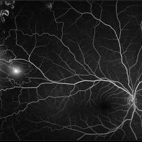 Proliferative Sickle Cell Retinopathy
Jan 29 2021 by Olivia Rainey
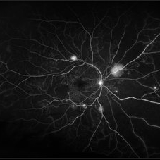
Ultra-widefield fluorescein angiogram of a 24-year-old female with proliferative sickle cell retinopathy affecting her right eye. The physician's interpretation of the fluorescein shows seafan neovascularization superotemporally, AV anastomeses, and good peripheral laser. He performed scatter PRP OD on 12/2/2020 to nonperfusion in temporal far periphery. The patient's 12/2020 Hb electrophoresis came back showing Hb SC (rather than sickle cell trait). Patient was born at full term, but she reports that her mother used drugs while pregnant with the patient. The patient also mentioned that her niece has full sickle cell disease and her grandmother, mother, and sibling all have condition on the sickle cell spectrum.
Photographer: Olivia Rainey, OCT-C, COA
Imaging device: Optos California
Condition/keywords: fluorescein angiogram (FA), fluorescein leakage, neovascularization (NV), neovascularization elsewhere (NVE), Optos, sea fan, sickle cell retinopathy
-
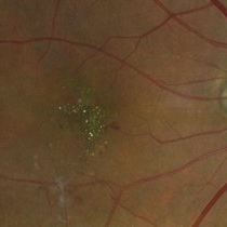 West African Crystalline Maculopathy
Oct 22 2023 by Niloofar Piri, MD
Fundus photograph of right eye of a patient from Liberia demonstrating multiple birefringent yellow green crystalline deposits in the fovea. Please notice the partially fibrosed NVE inferiorly. The disease has been shown to be associated with vascular disorders including diabetic retinopathy.
Photographer: Niloofar Piri, MD
Condition/keywords: crystalline maculopathy, crystalline retinopathy, West African Crystalline maculopathy
-
 Advanced Active PDR
Mar 29 2013 by Henry J. Kaplan, MD
Extensive NVD-FPD and NVE-FPE in a diabetic patient.
Condition/keywords: foveal photoreceptor defect, FPE, neovascularization (NV), neovascularization of the disc (NVD)
-
 Advanced PDR
Mar 29 2013 by Henry J. Kaplan, MD
Large FPD, FPE, NVE and multiple boat shaped subhyaloid hemorrhages inferiorly, ischemic retina and multiple occluded sclerotic vessels.
Condition/keywords: foveal photoreceptor defect, subhyaloid hemorrhage
-
 Branch Retinal Vein Occlusion
Dec 9 2020 by Olivia Rainey
Ultra-widefield angiogram of a 78-year-old male with a branch retinal vein occlusion affecting his right eye. The patient was diagnosed on 12/17/12 at another practice. The physician noted that there wasn't NVE noted, however areas of micoaneurysmal dilation is present. She noted retinal ischemia secondary to BRVO. 12/8/20 leakage on FA noted to be worsening compared to his previous angiography. She has concern for progressing NVE and recommends sector PRP after injection of antiVEGF series of 3 for the health of the eye.
Photographer: Olivia Rainey, OCT-C, COA
Imaging device: Optos California
Condition/keywords: branch retinal vein occlusion (BRVO), macular branch retinal vein occlusion (BRVO), non-perfusion, scleral buckle, vitreoretinal surgery
-
 Cheese Pizza Pie Appearance in CMV Retinitis
Mar 30 2024 by KANWALJEET HARJOT MADAN, M.S. (Ophthalmology); FAICO (Vitreous - Retina)
This is Fundus Photograph of left eye of 53 year male depicting an area of Retinal Necrosis with few Retinal Haemorrhages suggestive of CMV Retinitis. Areas of Perivascular Exudation also seen. On investigations, the patient was found to be HIV positive. He was started on Anti Retro Viral treatment after physician opinion.
Photographer: Dr. Kanwaljeet Harjot Madan, Thind Eye Hospital, Jalandhar City (Punjab) INDIA.
Imaging device: Zeiss Fundus Camera
Condition/keywords: AIDS, cytomegalovirus (CMV), retinitis
-
 Detached NVE During PVD induction
Apr 27 2018 by Michael J. Koss, MD, PhD, MBA
A 73-year-old woman with macular pucker underwent a pars plana vitrectomy with membrane peeling. Additionally the patient suffers from diabetic retinopathy after being diagnosed with type 2 diabetes mellitus sixteen years ago. Prior to the procedure she was treated with a series of intravitreal Bevacizumab-injections due to diabetic macular edema. There was no history of a proliferative DRP. During the vitrectomy a branch of an obliterated NVE spontaneously detached and floated freely in the vitreous. The 3D shot was captured via Alcon’s NGENUITY® 3D Visualization System in form of photograph and video providing an outstandingly detailed image of the branched NVE.
Photographer: Michael Koss, Augenzentrum Nymphenburger Hoefe
Imaging device: Alcon’s NGENUITY® 3D Visualization System
Condition/keywords: diabetes, diabetic retinopathy, neovascularization elsewhere (NVE), pars plana vitrectomy (PPV), PVD induction
-
 Diabetic Macular Edema, Proliferative Diabetic Retinopathy, Neovascularization Elsewhere, DME, PDR, NVE
Apr 1 2013 by James B. Soque, CRA, OCT-C, COA, FOPS
39-year-old white female and long standing diabetis, c/o new peripheral symptoms of left eye. FA OS reveals diabetic macular edema, microaneurysms, and neovasculaization elsewhere. Fluorescein Angogram, Early Phase, 50 Deg, 2x Mag.
Photographer: James B Soque, CRA, COA
Imaging device: Topcon TRC 50DX with MERGE software, OIS 10.6.45
Condition/keywords: diabetic macular edema, neovascularization (NV), proliferative diabetic retinopathy (PDR)
-
 Elevated Lesion
Nov 9 2012 by Norman Byer
This photograph and the next are two views of a very interesting elevated lesion in a 45-year-old man. This photograph shows the immense value of closely scrutinizing the profile of the indented area. Note that in the middle of the slide there is a sudden break in the continuity of the dark convex shadow that lies just behind the crest of the scleral indentation. If the elevated tissue is "filmy" or "wispy" or filamentous as in this case, it raises a strong suspicion that a retinal break is present just behind it.
Condition/keywords: elevated retinal lesion, elevated tissue, retinal break, scleral indentation
-
 Gyrate Atrophy
Feb 5 2024 by Ali Al-Ani, M.B.Ch.B, FRCS, FRCOphth, FAAO
A 22-year old woman presenting with a 3 year history of nyctalopia. Investigations showed elevated ornithine levels in plasma, urine and CSF.
Imaging device: OPTOS
Condition/keywords: gyrate atrophy
-
 Hypertensive Retinopathy
Dec 24 2017 by Purva Patwari
52-year-old female diagnosed of hypertension by retina evaluation.
Photographer: Dr Purva Patwari, Patwari Retina Center, Ahmedabad, Gujarat , India
Imaging device: ZEISS VISU500
Condition/keywords: hypertensive retinopathy, neovascularization elsewhere (NVE), Roth spots
-
 Multifocal Pattern Dystrophy
Feb 5 2025 by Kimberly Wakester
Optomap RGB and AF photograph of an 37-year-old woman with multifocal pattern dystrophy in both eyes. Previously believed to be Stargardts, but genetic testing returned positive for PRPH2 mutation. Likely Multifocal Pattern Dystrophy given phenotypical appearance of SGD. There is stable NVE in the left eye. Will continue to monitor both eyes and consider treatment with PRP laser if needed for NVE in the left eye.
Photographer: Kimberly Wakester, COA
Imaging device: Optos California
Condition/keywords: multifocal pattern dystrophy, NVE, PRPH2 Positive
-
 NEOVASCULARISATION OF DISC- OCT-ANGIOGRAPHY
Jun 13 2023 by Sonali P Lomte, MBBS,DNB
OCT Angiography of Optic Disc ( vitreous slab) of a 56 year old male with proliferative diabetic retinopathy showing neovascularization of disc.
Photographer: Dr Sonali Lomte, R J Sankara Eye Hospital, New Panvel
Imaging device: TOPCON DRI OCT Triton Plus swept source OCT
Condition/keywords: NEOVASCULARISATION OF DISC, OCTA
-
 NVE in a Patient With Vasculitis
Nov 5 2018 by awaneesh m upadhyay, MBBS, DNB
FFA image of a 22-year-old male vasculitis patient with NVE.
Photographer: Hiteshwar Saikia
Condition/keywords: neovascularization elsewhere (NVE), tuberculosis, vasculitis
-
 Proliferative Diabetic Retinopathy
Jan 29 2021 by Olivia Rainey
Ultra-widefield fluorescein angiogram of a 65-year-old male with proliferative diabetic retinopathy affecting his right eye. The patient's diabetic retinopathy has progressed significantly since he was last seen in 2014. It was recommended to begin antiVEGF to control DME followed by laser treatment OU.
Photographer: Olivia Rainey, OCT-C, COA
Imaging device: Optos California
Condition/keywords: anti-VEGF, diabetes, diabetic macular edema, fluorescein angiogram (FA), fluorescein leakage, neovascularization (NV), neovascularization elsewhere (NVE), non-perfusion, Optos, proliferative diabetic retinopathy (PDR), ultra-wide field imaging
-
 Proliferative Diabetic Retinopathy
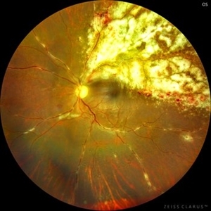
May 24 2024 by Anjana Mirajkar, MS Ophthalmology
A central photo of a 65 year old female of right eye showing fibro vascular proliferation with neovascularization elsewhere in a case of proliferative diabetic retinopathy.
Photographer: Dr. Anjana Mirajkar -Retina Foundation, Ahmedabad
Imaging device: Mirante-Nidek
Condition/keywords: NVE, proliferative diabetic retinopathy (PDR), tractional retinal detachment
-
 Proliferative Diabetic Retinopathy with Macular Hole IVFA
Jul 26 2020 by Gareth Lema, MD, PhD
IVFA shows NVE along the inferotemporal arcade with ischemia immediately inferior to the NV.
Condition/keywords: macular hole, proliferative diabetic retinopathy (PDR)
-
 ROP 4B late Retinal Findings
Mar 31 2022 by Franco Benvenuto, MD
A 9-year-old male, that was born at 30 weeks of gestation with birth weight of 1500 g and history of hospitalization for 20 days with respiratory distress and packed red blood cell transfusion for anemia. At the first exam, both eyes were with stage 4B ROP. Vitrectomy with 25 G was done in both eyes. The flat fibrosis dragged the macula nasally in both the eyes.
Photographer: Franco Benvenuto, Universidad de Buenos Aires, Argentina; Universidad de Guadalajara, México.
Condition/keywords: cicatricial retinopathy of prematurity, retinopathy of prematurity (ROP)
-
 Subhyaloid Hemorrhage with JXT and Proliferative Diabetic Retinopathy
Jan 13 2022 by ASRS Staff
Wide field photograph of 50 year-old female, known case of idiopathic juxtafoveal telangiectasia in both eyes and known diabetic, presented with subhyaloid hemorrhage and NVE.
Imaging device: Nidek Mirante
Condition/keywords: florid type PDR, JXT, neovascularization elsewhere (NVE), subhyaloid hemorrhage, ultra-wide field imaging
-
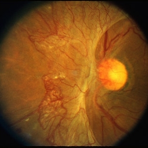
 Superior Hemi-Central Retinal Artery Occlusion
Apr 24 2024 by Mosab Salah
Fundus photograph -inverted view- taken by smartphone fundus photography, of a young man with sudden onset altitudinal field defect, a Superior Hemi-Central Retinal Artery Occlusion noted.
Photographer: Dr Mosab Salah, The Islamic Hospital, Amman, Jordan
Imaging device: smartphone fundus photography and 30 D Lens
Condition/keywords: arterial occlusion, branch retinal artery occlusion (BRAO), BRAO, CRAO, Hemi-Central Retinal Artery Occlusion (CRAO), occlusive vasculitis, smartphone fundus photography
-
 Tractional Retinal Detachment
Apr 20 2024 by Tejaswita Verma
Fundus photograph of the right eye of a 62 year old female with tractional retinal detachment in a case of lasered proliferative diabetic retinopathy showing neovascularisation at disc and elsewhere
Photographer: DR. TEJASWITA VERMA
Condition/keywords: Neovascularisation at the Disc (NVD), Neovascularisation elsewhere (NVE), proliferative diabetic retinopathy (PDR)
 Loading…