Search results (264 results)
-
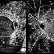
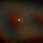
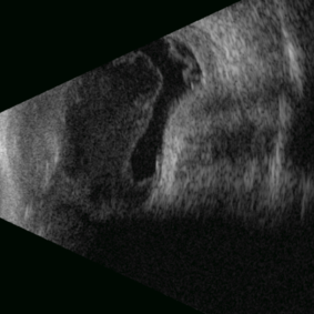 Posterior Nodular Scleritis
Apr 23 2025 by Gustavo Uriel Fonseca Aguirre
This B-mode ultrasound scan demonstrates a posterior scleral nodule accompanied by vitritis, serous retinal detachment, and annular choroidal detachment. The nodule appears as a localized hypoechoic scleral thickening, while the serous retinal detachment shows a smooth convex configuration. The choroidal detachment presents with the characteristic ring-shaped elevation, suggesting significant intraocular inflammation or hypotony.
Photographer: Gustavo U. Fonseca Aguirre, Hospital Conde de Valenciana, Ciudad de México
Condition/keywords: posterior nodular scleritis, posterior scleritis
-
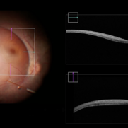
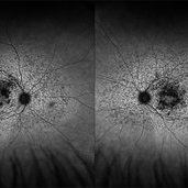
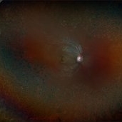
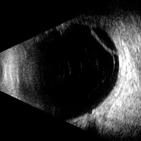 Retinoschisis
Apr 21 2025 by Gustavo Uriel Fonseca Aguirre
This B-mode longitudinal ultrasound scan reveals a peripheral temporal retinoschisis, demonstrating a characteristic thin, dome-shaped separation of the retinal layers without associated subretinal fluid or vitreous traction. The lesion shows smooth, convex contours with maintained structural integrity of both retinal layers.
Photographer: Gustavo U. Fonseca Aguirre, Hospital Conde de Valenciana, Ciudad de México
Condition/keywords: retinoschisis
-
Eye of the Hurricane
Apr 9 2025 by Gustavo Uriel Fonseca Aguirre
Ultrasound biomicroscopy of a post-operative eye (status post trabeculectomy and phacoemulsification) reveals a patent ostium on the right side, along with an intraocular lens in position. A hyphema is observed displaying small convection currents, creating a circular motion pattern due to the temperature gradient between the iris and cornea. Notably, the blood flow can be seen circulating toward the trabeculectomy site.
Condition/keywords: hyphema, trabeculectomy
-
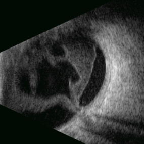
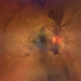
 Eye of the Hurricane
Apr 8 2025 by Gustavo Uriel Fonseca Aguirre
Ultrasound biomicroscopy of a post-operative eye (status post trabeculectomy and phacoemulsification) reveals a patent ostium on the right side, along with an intraocular lens in position. A hyphema is observed displaying small convection currents, creating a circular motion pattern due to the temperature gradient between the iris and cornea. Notably, the blood flow can be seen circulating toward the trabeculectomy site.
Photographer: Gustavo U. Fonseca Aguirre, Hospital Conde de Valenciana, Ciudad de México
Condition/keywords: Hyphema, trabeculectomy
-
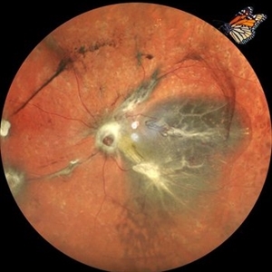
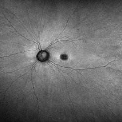
 Stag Horn
Apr 8 2025 by Gustavo Uriel Fonseca Aguirre
B-mode ultrasound of a young male patient with bilateral panuveitis (currently under investigation) reveals intense vitritis with islands of preserved vitreous and partial posterior hyaloid detachment, creating a characteristic "stag horn" appearance.
Photographer: Gustavo U. Fonseca Aguirre, Hospital Conde de Valenciana, Ciudad de México
Condition/keywords: Panuveitis
-
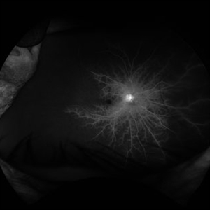
 Branches Starved of Flow, Yet Nature Strives to Grow
Apr 1 2025 by rohan jain
Tufts of NVE's in a case of Branch Retinal Vein Occlusion
Photographer: Dr. ROHAN JAIN
Condition/keywords: branch retinal vein occlusion (BRVO), capillary nonperfusion, non-perfused branch retinal vein occlusion (BRVO), non-perfusion, NVE, OCT Angiography, ST BRVO
-
 Actively Bleeding NVE
Apr 1 2025 by Jordyn Beckman
47 year old woman presented with actively bleeding NVE temporally on exam with complaints of foggy vision and floaters.
Photographer: Jordyn Beckman, Retina Consultants of Carolina, P.A.
Imaging device: Optos California
Condition/keywords: active bleeding, Elevated retinal neovascularization, vitreous hemorrhage
-
 Completed Bleb with OCT Through Fovea
Mar 25 2025 by Robert Andrew Sisk, MD, FACS, FASRS
Color still from surgical video of subretinal delivery of laru-zova for X-linked retinitis pigmentosa. Live optical coherence tomography (OCT) with foveal tracking via the embedded software in the operating microscope allows monitoring foveal integrity for signs of stress. The contour of the fovea does not exceed the curvature of the bleb (e.g. no inversion). The tangential cannula angle facilitated steering of the bleb posteriorly. The bleb covers essentially the entire macula, which is the target area.
Imaging device: Zeiss Artevo 800
Condition/keywords: gene therapy, genetic disorder, optical coherence tomography (OCT), retinitis pigmentosa, subretinal injection
-
 Combined Retinal Detachment With a Butterfly Shaped Configuration
Mar 13 2025 by S. Natarajan, MD, FASRS, FRCS (GLASGOW) , FICO, D.Sc, FELA
A 46 year old female presented to us with diminished vision in both the eyes. Her blood glucose levels were deranged. She had bilateral proliferative diabetic retinopathy and pan retinal photocoagulation was done elsewhere. Left eye showed a combined retinal detachment with fibrovascular proliferation on the disc and along inferior arcade with a convex configuration of retinal detachment. Patient was planned for surgical intervention. The image shows a butterfly like configuration of combined retinal detachment with the subretinal fluid pocket appearing like the wings of the butterfly.
Photographer: ASHWINI SUTAR ADITYA JYOT EYE HOSPITAL
Imaging device: Mirante ( PLEASE SELECT COVER PAGE )
Condition/keywords: retinal detachment with a butterfly shaped
-
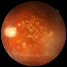 Emulsified Silicon Oil in Macular Hole- Hyperoleon in Hole
Mar 8 2025 by PUJA NEGI
Fundus photograph showing emulsified silicon oil bubbles in macular hole, hyperoleon in hole.
Photographer: Dr Vaidehi Jethwa
Condition/keywords: Inverse hypopyon, macular hole
-
 Proliferative Diabetic Retinopathy S/P Pan Retinal Photocoagulation
Mar 4 2025 by Prithvi Chandrakanth
A 52-year-old female patient presented with complaints of diminishing vision, compounded by uncontrolled diabetes mellitus. Her Fundus examination revealed proliferative diabetic retinopathy, characterized by neovascularization of the disc and elsewhere, and sclerosed vessels. To address this, Pan Retinal Photocoagulation was performed, and the condition stabilized, halting the progression of the disease.
Photographer: DR PRITHVI CHANDRAKANTH, DR CHANDRAKANTH NETHRALAYA, KOZHIKODE, KERALA, INDIA
Imaging device: EIDON
Condition/keywords: Diabetic Retinopathy, Neovascularisation at the Disc (NVD), neovascularization of the disc (NVD), NVD, pan-retinal photocoagulation (PRP), PDR, PDR with NVE (periphery), PRP
-
 Bilateral Proliferative Diabetic Retinopathy OU
Feb 21 2025 by Drew Mitchell
OCT-Angiography 8x8 Montage OU. PDR with active NVE OD. 37 year old male with no visual complaints. Vision is 20/20 in both eyes.
Photographer: Drew Mitchell OCT-C
Imaging device: Zeiss Cirrus 5000
Condition/keywords: CIRRUS 5000 ANGIOPLEX, Diabetes, NVE, OCT Angiography, proliferative diabetic retinopathy (PDR)
-
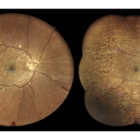
 Multifocal Pattern Dystrophy
Feb 5 2025 by Kimberly Wakester
Optomap RGB and AF photograph of an 37-year-old woman with multifocal pattern dystrophy in both eyes. Previously believed to be Stargardts, but genetic testing returned positive for PRPH2 mutation. Likely Multifocal Pattern Dystrophy given phenotypical appearance of SGD. There is stable NVE in the left eye. Will continue to monitor both eyes and consider treatment with PRP laser if needed for NVE in the left eye.
Photographer: Kimberly Wakester, COA
Imaging device: Optos California
Condition/keywords: multifocal pattern dystrophy, NVE, PRPH2 Positive
-
 Eales Disease
Jan 31 2025 by Thirumalesh Mochi Basavaraj, MD
Ultra wide field image of a 24 year-old young healthy adult male with a visible sea fan neovascularization with partial PVD secondary to Scatter LASER photocoagulation with Vitreous and subhyaloid hemorrhage.
Photographer: Puttaswamy N K
Condition/keywords: Eales disease, Neovascularisation elsewhere (NVE), sea fan
-
Macular Hole Surgery: Inverse Flaps
Jan 31 2025 by Thirumalesh Mochi Basavaraj, MD
This video demonstrates, PVD induction , followed by ILM peeling in multiple flower petal flap technique.
Condition/keywords: ILM flaps, ILM peel, induction
-
 Systemic Lupus Erythematosus (SLE) Vasculitis
Jan 29 2025 by Kimberly Wakester
Fundus photographs of an 13-year-old boy with Systemic Lupus Erythematosus (SLE) Vasculitis in both eyes s/p PRP laser. Patient is doing well s/p PRP Laser OU and with continued use of oral medications. Patient will be monitored with follow up exams to check for recurring vasculitis or recurring/worsening NVE/NVD. Patient is to continue ongoing management with Rheumatologist.
Photographer: Kimberly Wakester, COA
Imaging device: Optos California
Condition/keywords: NVD, NVE, occlusive vasculitis, pan-retinal photocoagulation (PRP), Systemic Lupus Erythematosus (SLE) Vasculitis
-
 Sickle-Cell Retinopathy
Jan 22 2025 by Virginia Gebhart
Fluorescein angiogram of 54 year old female with non-diabetic proliferative retinopathy. Recent labs confirm sickle-cell disease. FA shows temporal peripheral non perfusion with NV. S/p PRP with retrobulbar block
Photographer: Virginia Gebhart, Retina Consultants of Carolina
Imaging device: Optos California
Condition/keywords: FA, Fluorescein angiography, Neovascularisation elsewhere (NVE), non-perfusion, Nose, pan-retinal photocoagulation (PRP), PRP, sickle cell retinopathy
-
 Raised NVD
Jan 9 2025 by Richa Chaudhary, Mbbs,ms
62 year old female, with h/o vein occlusion, presented with florid raised NVE , regressed well with intravitreal anti-VEGF.
Condition/keywords: neovascularization of the disc (NVD)
-
 Dome Shaped Macula
Jan 7 2025 by Jordyn Beckman
Autofluorescence photograph of 36 year old woman with Dome Shaped Macula with hypoautofluorescence and hyperautofluorescence centrally.
Photographer: Jordyn Beckman
Imaging device: California Optos
Condition/keywords: autoflorescence, convex protrusion, dome shaped macula, high myopia, hyperautofluorescent centrally, hypoautofluorescence
-
 Treatment of Ocular Ischemic Syndrome with Hyperbaric Oxygen Therapy
Dec 2 2024 by Catherine S Kang
A 66-year-old female with past medical history significant for hypertension and ocular ischemic syndrome. She presented in emergency department (ED) reporting eye pain and blurred vision in both eyes since earlier that morning. On examination, best corrected visual acuity in each eye was counting fingers (20cm). Further investigation was performed and fluorescein angiography revealed a delay in choroidal filling. The patient has been followed for ocular ischemic syndrome since the onset of the condition and hyperbaric oxygen therapy was promptly initiated. Final best corrected visual acuity was 20/150 and macula developed atrophy.
Photographer: Catherine Kang
Condition/keywords: hyperbaric oxygen therapy, ocular ischemic syndrome
-
 Proliferative Diabetic Retinopathy with Neovascularization
Nov 15 2024 by Júlio Andrade
Fundus photograph of a patient with proliferative diabetic retinopathy presenting with extensive neovascularization, microaneurysms, and evidence of fluid leakage. Key findings include the presence of new blood vessel formation, indicative of advanced retinal ischemia and disease progression.
Photographer: úlio Andrade, Retina Instituto, Belo Horizonte
Imaging device: Zeiss Clarus 700
Condition/keywords: diabetic blindness, ischaemic diabetic maculopathy, Neovascularisation elsewhere (NVE), NEOVASCULARISATION OF DISC, Proliferative Diabetic retinopathy
-
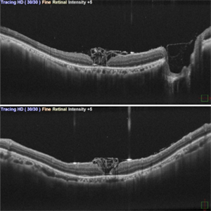 Inverted ILM Flap in RD with FTMH-First Post Op Day
Oct 29 2024 by SHILPI H NARNAWARE, ICO ( Retina) , FAICO ( Vitreo-Retina)
OCT showing stuffed ILM under oil on first post op day in a case of RRD with FTMH.
Photographer: Shilpi Narnaware, Sarakshi Netralaya , Nagpur, Maharashtra , India
Imaging device: Mirante ( by Nidek)
Condition/keywords: FTMH, Inverted ILM technique, RD
 Loading…