Search results (403 results)
-
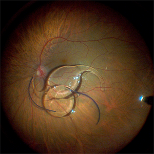
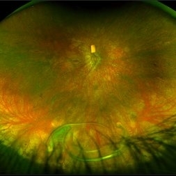
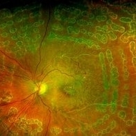
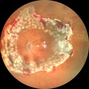 360 Degree Retinectomy
Sep 11 2020 by Sham Talati, DOMS
A case of retinal detachment with PVR. Patient underwent pars plana vitrectomy with silicon oil injection with 360 degree retinectomy.
Photographer: Dr. Sham Talati,Retina Foundation,Ahmedabad
Imaging device: Nidek Mirante
Condition/keywords: proliferative vitreoretinopathy (PVR), retinectomy
-
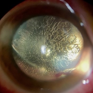 Feather like cataract
Apr 11 2023 by rodrigo torres
Cataract after vitrectomy and gas tamponade.
Photographer: Rodrigo Amaral Torres
Condition/keywords: cataract, pars plana vitrectomy (PPV)
-
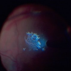
 Seedlings of Fungal Endophthalmitis
Mar 14 2025 by SHILPI H NARNAWARE, ICO ( Retina) , FAICO ( Vitreo-Retina)
57 year diabetic female , was treated as a case of recurrent vitreous post cataract surgery. Patient was posted for vitrectomy 3 months post cataract surgery. Intra-operatively, multiple yellowish colonies were seen all over the posterior pole were seen, which were later found to be Aspergillus colonies.
Photographer: Shilpi Narnaware, Sarakshi Netralaya , Nagpur, Maharashtra , India
Imaging device: Ngenuity
Condition/keywords: endophthalmitis, fungal
-
 "Internal Mirroring" Effect by Intraocular Gas
Mar 25 2014 by Homayoun Tabandeh, MD, FASRS
"Internal mirroring" by residual intraocular gas in a highly myopic patient 3 weeks post repair of retinal detachment with pars plana vitrectomy and C3F8 gas.
Photographer: Danny Rivas
Condition/keywords: high myopia, intraocular gas
-
 Lady in a dress
Feb 9 2023 by Shelby Helton
Fluorescein Angiography on a 67-year-old male with significant RPE changes secondary to a severe subretinal hemorrhage that required a vitrectomy with subretinal TPA in 2013.
Photographer: Shelby Helton
Imaging device: Heidelberg Spectralis
Condition/keywords: wet age-related macular degeneration (wet AMD)
-
 Fraternal Twins
May 22 2023 by Gustavo M. Hüning, MD, MBA, FASRS
Intrasurgical photograph using a non-contact system and 3D visualization system of a 65-year-old woman who suffered an ocular trauma.
Photographer: Gustavo M. Hüning, Hüning Clínica do Olhar, Santa Maria - Brazil
Imaging device: Alcon Luxor combined with Alcon nGenuity
Condition/keywords: dislocated intraocular lens (IOL), implant, pars plana vitrectomy (PPV)
-
 Intraoperative Photo Taken During Vitrectomy
Jan 26 2017 by Manish Nagpal, MD, FRCS (UK), FASRS
Intraoperative photo while doing vitectomy near a horseshoe tear to clear the adherent vitreous enhanced by peripheral scleral indentation while using chandelier light.
Photographer: Manish Nagpal
Imaging device: Still captured from a 3 chip HD camera on microscope
Condition/keywords: cutter, scleral indentation, vitrectomy, vitreous
-
 Post Subretinal tpa , viterectomy and gas
May 6 2022 by Shobhit Chawla, M.S.
SUBMACULAR HAEMORRHAGE IN A 38YEAR OLD LADY PATIENT CAUSE POLYP BLEED IN PCV. Following viterectomy , subretinal tpa . gas and aflibercept injection. 7 day post operative image.
Photographer: Shobhit Chawla
Imaging device: Zeiss Clarus 500
Condition/keywords: aflibercept, intravitreal gas bubble, submacular hemorrhage, tissue plasminogen activator (tPA), vitrectomy
-
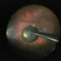 PPV retained cataract
Apr 19 2023 by Denica Rodriguez
A 46-year-old male with hypermature dense cataract. Patient got a piece of metal in his eye when he was 5 years old and was not able to see since. Patient was having cataract surgery and phacodonesis was present. The lens dropped to the back of the eye. Patient had to have another surgery to do vitrectomy. The lens removal was done with a fragmatome handpiece.
Photographer: Denica Rodriguez COA, ST
Imaging device: Zeiss Microscope with resight
Condition/keywords: cataract, dropped nucleus, fragmatome, lens capsule, ocular trauma, pars plana vitrectomy (PPV), retained lens fragments, Retina, retina surgery, traumatic cataract
-
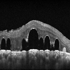
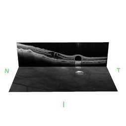 Retained Perfluorcarbon and Macular Edema After Silicon Oil Removal 3D
Jul 24 2017 by Nelson Chamma Capelanes, MD
SD-OCT and HRA from a 42-year-old patient after silicon oil removal. The image shows macular edema and retained perfluorcarbon.
Photographer: Nelson Chamma Capelanes, Promedica Indaiatuba, Brazil
Condition/keywords: macular edema, post-vitrectomy, retained perfluorocarbon
-
 submacular perfluorocarbon liquid
Sep 7 2022 by JEFFERSON R SOUSA, Tecg.º (Biomedical Systems Technology)
A 63-year-old male patient underwent vitreoretinal surgery with the use of perfluorocarbon. From a technological point of view, extended-field retinography presents many points of focus variation due to the difficulty of establishing a diffuse focus, as it is a recent post-operative case. In OCT Fundus Enface, although it has a low resolution, it is extremely important for documenting the presence of perfluor. Best seen in structural OCT.
Photographer: JEFFERSON ROCHA DE SOUSA - Retinal Department at Instituto Dr. Suel Abujamra Sao Paulo-Brazil
Imaging device: Optical Coherence Tomography system OCT CIRRUS 5000, Protocol, HD 5 Line
Condition/keywords: perfluorocarbon fluid, post-vitrectomy, submacular perfluorocarbon liquid (PFO), vitrectomy
-
000---thumb.jpg/image-square;max$300,300.ImageHandler) Anterior Segment Photo of Emulsified Silicone Oil
Anterior Segment Photo of Emulsified Silicone Oil
Dec 25 2013 by Dong Yoon Kim, MD
47-year-old woman underwent vitrectomy and silicone oil tampoande for tractional retinal detachment due to proliferative diabetic retinopathy. 8 months after silicone oil tamponade, silicone oil was emulsified. And emulsified silicone oil was observed at anterior chamber.
Condition/keywords: silicone oil, tractional retinal detachment
-
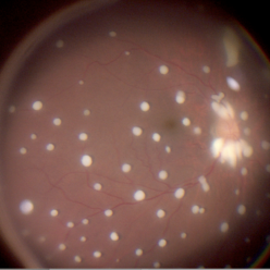
---thumb.jpg/image-square;max$300,300.ImageHandler) C3F8 gas bubble after retinal detachment surgery
C3F8 gas bubble after retinal detachment surgery
Feb 1 2013 by Sharon Fekrat, MD FACS FASRS
63 year old man s/p encircling scleral buckle and 23g pars plana vitrectomy for a macula off phakic rhegmatogenous retinal detachment. This fundus photograph shows the effect of the encircling buckle and the residual C3F8 intravitreal gas bubble in the right eye.
Photographer: Tiffanie Keaton, Duke Eye Imaging, Duke University Eye Center, Durham, NC
Imaging device: Optos
Condition/keywords: intravitreal gas bubble, vitrectomy
-
 Chronical Submacular Hemorrhage in the Setting of Neovascular AMD
Mar 23 2015 by Rita Couceiro, MD, MS
An 80-year-old male, with a history of hypertension and high cholesterol, complained of acute and painless vision loss in his left eye (OS) in the previous 5 months. On observation best corrected visual acuity in OS was hand motion. A dense vitreous opacity in OS precluded fundus examination. Ocular ultrasound revealed vitreous hemorrhage and thickening of the macular area. The patient was submitted to pars plana vitrectomy, which disclosed a large submacular hemorrhage with chronical features and disciform scarring in the setting of neovascular AMD.
Imaging device: Intraoperative fundus photograph
Condition/keywords: neovascular age-related macular degeneration (AMD), submacular hemorrhage, wet age-related macular degeneration (wet AMD)
-
 Detached NVE During PVD induction
Apr 27 2018 by Michael J. Koss, MD, PhD, MBA
A 73-year-old woman with macular pucker underwent a pars plana vitrectomy with membrane peeling. Additionally the patient suffers from diabetic retinopathy after being diagnosed with type 2 diabetes mellitus sixteen years ago. Prior to the procedure she was treated with a series of intravitreal Bevacizumab-injections due to diabetic macular edema. There was no history of a proliferative DRP. During the vitrectomy a branch of an obliterated NVE spontaneously detached and floated freely in the vitreous. The 3D shot was captured via Alcon’s NGENUITY® 3D Visualization System in form of photograph and video providing an outstandingly detailed image of the branched NVE.
Photographer: Michael Koss, Augenzentrum Nymphenburger Hoefe
Imaging device: Alcon’s NGENUITY® 3D Visualization System
Condition/keywords: diabetes, diabetic retinopathy, neovascularization elsewhere (NVE), pars plana vitrectomy (PPV), PVD induction
-
 Dexamethasone Implant
Jul 3 2021 by Gerardo Rivera Arroyo
42-year-old male, operated on for vitrectomy plus scleral buckling plus silicone plus dexamethasone implant for inferior retinal detachment with PVR.
Photographer: Rosa Elizabeth Moreno Anda, MD, Hospital Central Militar CDMX
Condition/keywords: dexamethasone implant, retina surgery, vitrectomy
-
 Dislocated Lens
Apr 26 2023 by Chloe Hanifan
Ultra wide field fundus photograph of a 41-year-old male with a dislocated lens affecting his right eye. IOL noted inferior vitreous base and vitrectomy surgery for removal of IOL was recommended. Patient has history of retinitis pigmentosa as well. Patient's vision at the time of presentation was counting fingers at 2 feet.
Photographer: Chloe Hanifan
Imaging device: Optos California
Condition/keywords: dislocated lens, fundus photography, Optos, pseudocolor, retinitis pigmentosa, ULTRA WIDE FIELD
-
 Dislocated Lens, Posterior OD
Jan 26 2024 by Corey Grant
OPTOS California photo presents a 71 year old male patient with a dislocated lens, posterior in the right eye. Presented on 1/26/24 with posteriorly dislocated SN60WF with a Soemmerring ring. Associated retinal hemorrhage within retinoschisis as well. This will result in a PPV/IOL exchange/SFIOL/STK for the right eye.
Photographer: Corey Grant, Ophthalmic Imager, Retina Specialist of Michigan
Imaging device: OPTOS California
Condition/keywords: color photo, IOL, OD, Optos, OPTOS CALIFORNIA, pars plana vitrectomy (PPV), retina
-
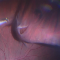
 Dislocation of the Crystalline Lens with a Retinal Detachment
Apr 21 2025 by Hrishikesh Naik, MS
An intraoperative screen grab shows a dislocation of the crystalline lens along with an associated rhegmatogenous retinal detachment in a case of Marfan’s syndrome. The case was managed by a combined PPV-SB procedure. A vitrectomy cutter is seen at the left.
Photographer: Hrishikesh Naik
Condition/keywords: intraoperative, lens dislocation, Marfan's syndrome, Retinal Detachment, vitrectomy
-
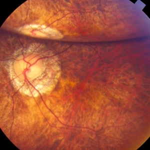
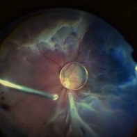
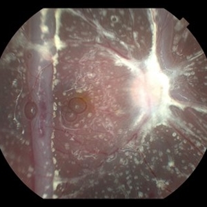
 Endolaser in Status-Post Vitrectomy
Aug 28 2023 by Aditya S Kelkar, MS, FRCS, FASRS,FRCOphth
Endolaser in Status-Post Vitrectomy.
Photographer: Optom Komal Jangam, National Institute of Ophthalmology, Pune, India.
Imaging device: OPTOS DAYTONA
Condition/keywords: endolaser, pars plana vitrectomy (PPV), vitrectomy
-
 Epiretinal Membrane
Oct 11 2012 by Michael P. Kelly, FOPS
This is a patient with idiopathic panuveitis who developed a visually significant epiretinal membrane. Pars plana vitrectomy with membrane peeling was performed to remove the epiretinal proliferation. I recommend magnifying the image to see the exquisite detail centrally.
Photographer: Michael P. Kelly, FOPS Director, Duke Eye Center Labs, Duke Universtiy Hospital
Imaging device: Zeiss 450Plus
Condition/keywords: epiretinal membrane (ERM), panuveitis
-
Epiretinal membrane removal
Oct 24 2022 by Manish Nagpal, MD, FRCS (UK), FASRS
This video highlights the surgical technique of tangentially removing the epiretinal membrane using a forceps
Photographer: Manish Nagpal
Condition/keywords: epiretinal membrane, ERM, macular pucker, staining, video, vitrectomy
-
 Exudative Macular Detachment After Intensive Laser Photocoagulation
Mar 12 2016 by Sjakon G Tahija, MD
Fundus photograph of 44-year-old man with exudative detachment of the macula after vitrectomy and ILM peeling for proliferative diabetic retinopathy combined with intensive endolaser photocagulation.
Photographer: Avris Siahaan, Klinik Mata Nusantara
Condition/keywords: exudative detachment, pan-retinal photocoagulation (PRP)
-
 Fish Hook Eye Trauma
Jun 12 2024 by Miguel Brito, MD, FASRS
Fundus photograph of a 15-year-old boy post cataract aspiration, pars plana vitrectomy, suprachoroidal drainage, and retinal reattachment surgery secondary to traumatic endophthalmitis.
Photographer: Miguel Brito
Condition/keywords: endophthalmitis, PFCL, Retinal detachment under Silicon Oil, retinal fold
-
Flattening a bullous retinal detachment
Oct 24 2022 by Manish Nagpal, MD, FRCS (UK), FASRS
This surgical clip shows the way a bullous retinal detachment reattaches at the time of endodrainage
Photographer: Manish Nagpal
Condition/keywords: bullous detachment, endodrainage, tear, video, vitrectomy
 Loading…