 Initializing download.
-
 By Michael J. Koss, MD, PhD, MBA
Co-author(s): Marie Isabel Schaller, Gregor Kastl, Quyhn-Trang Ippisch, Peter Heidenkummer - Uploaded on Apr 27, 2018.
- Last modified by Caroline Bozell on May 1, 2018.
- Rating
- Appears in
- Miscellaneous
- Condition/keywords
- diabetic retinopathy, neovascularization elsewhere (NVE), pars plana vitrectomy (PPV), PVD induction, diabetes
- Photographer
- Michael Koss, Augenzentrum Nymphenburger Hoefe
- Imaging device
- Alcon’s NGENUITY® 3D Visualization System
- Description
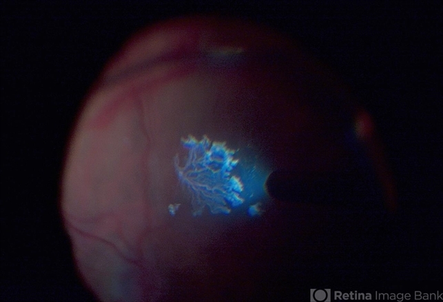
- A 73-year-old woman with macular pucker underwent a pars plana vitrectomy with membrane peeling. Additionally the patient suffers from diabetic retinopathy after being diagnosed with type 2 diabetes mellitus sixteen years ago. Prior to the procedure she was treated with a series of intravitreal Bevacizumab-injections due to diabetic macular edema. There was no history of a proliferative DRP. During the vitrectomy a branch of an obliterated NVE spontaneously detached and floated freely in the vitreous. The 3D shot was captured via Alcon’s NGENUITY® 3D Visualization System in form of photograph and video providing an outstandingly detailed image of the branched NVE.


