Search results (403 results)
-
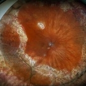
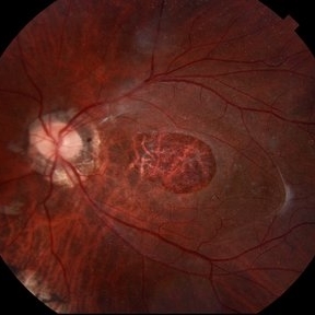
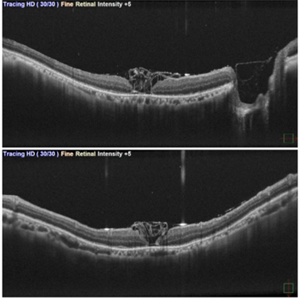
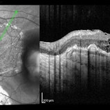
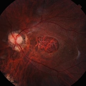 Giant Persistent Macular Hole
Jul 5 2025 by César Adrián Gómez Valdivia, MD
Giant Persistent Macular Hole found in a 48YO male patient 1 year after vitrectomy.
Photographer: @eyemissu2
Imaging device: TOPCON TRC-50DX
Condition/keywords: macular hole
-
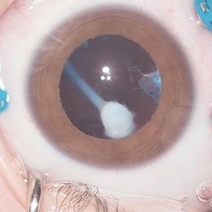
 Dislocated Cataractous Lens
Jun 19 2025 by Mrinali Gupta, MD, FASRS
Intraoperative image of a chronically dislocated cataractous lens. The patient underwent pars plana vitrectomy, lensectomy, and placement of an anterior chamber intraocular lens, with improvement in vision from Count Fingers to 20/20 without correction.
Photographer: Mrinali Gupta, MD
Imaging device: Intraoperative surgical video (Zeiss Lumera scope, Resight lens)
Condition/keywords: dislocated crystalline lens
-
 Dislocation of the Crystalline Lens with a Retinal Detachment
Apr 21 2025 by Hrishikesh Naik, MS
An intraoperative screen grab shows a dislocation of the crystalline lens along with an associated rhegmatogenous retinal detachment in a case of Marfan’s syndrome. The case was managed by a combined PPV-SB procedure. A vitrectomy cutter is seen at the left.
Photographer: Hrishikesh Naik
Condition/keywords: intraoperative, lens dislocation, Marfan's syndrome, Retinal Detachment, vitrectomy
-
 Proliferative Vitreoretinopathy
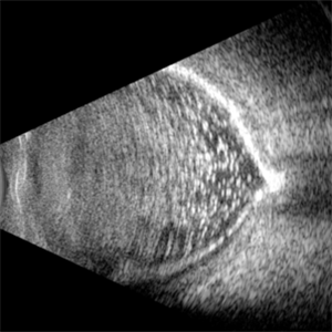
Apr 17 2025 by Gustavo Uriel Fonseca Aguirre
This B-mode transverse ultrasound scan depicts a post-vitrectomy eye with recurrent retinal detachment in a patient with diabetic retinopathy history. The image reveals fresh vitreous cavity hemorrhage and subretinal bleeding, along with subretinal proliferative bands (PVR strands). These findings indicate complicated tractional re-detachment with active hemorrhagic components.
Photographer: Gustavo U. Fonseca Aguirre, Hospital Conde de Valenciana, Ciudad de México
Condition/keywords: proliferative vitreoretinopathy (PVR)
-
 Calcification of the Retina
Apr 7 2025 by Gustavo Uriel Fonseca Aguirre
B-mode ultrasound of a vitrectomized eye reveals emulsified silicone oil in the vitreous cavity, retinal detachment, and calcification of the retina and optic nerve head.
Photographer: Gustavo U. Fonseca Aguirre, Hospital Conde de Valenciana, Ciudad de México
Condition/keywords: calcification, Retina detachment, vitrectomy
-
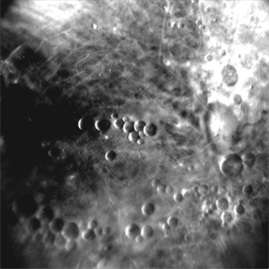
 Vasoproliferative Tumor
Mar 25 2025 by Gustavo Uriel Fonseca Aguirre
Patient diagnosed with pars planitis and a history of phacovitrectomy. Longitudinal B-scan section showing a very pronounced, homogeneous tumor lesion in the periphery. The A-scan revealed high average reflectivity with an irregular internal structure.
Photographer: Gustavo U. Fonseca Aguirre, Hospital Oftalmológico de la Luz, Ciudad de México
Condition/keywords: pars planitis, Vasoproliferative Tumor
-
 Repaired Retinal Detachment with PVR
Mar 25 2025 by Kimberly Wakester
Optomap RGB of a 79-year-old-woman with a repaired retinal detachment with PVR in the right eye. Patient is doing well over 7 months s/p vitrectomy with silicone oil and scleral buckle placement. Retina remains attached on the buckle under oil. Patient is to return in 6 months for follow up exam with repeat imaging.
Photographer: Kimberly Wakester, COA, OCT-C
Imaging device: Optos California
Condition/keywords: PVR, repaired RD, Retinal detachment under Silicon Oil, scleral buckle
-
 Seedlings of Fungal Endophthalmitis
Mar 14 2025 by SHILPI H NARNAWARE, ICO ( Retina) , FAICO ( Vitreo-Retina)
57 year diabetic female , was treated as a case of recurrent vitreous post cataract surgery. Patient was posted for vitrectomy 3 months post cataract surgery. Intra-operatively, multiple yellowish colonies were seen all over the posterior pole were seen, which were later found to be Aspergillus colonies.
Photographer: Shilpi Narnaware, Sarakshi Netralaya , Nagpur, Maharashtra , India
Imaging device: Ngenuity
Condition/keywords: endophthalmitis, fungal
-
 Inadvert Globe Perfuration After Peribulbar Block
Mar 13 2025 by Bruno B Ribeiro
Fundus photograph of a 74-year-old woman who underwent pars plana vitrectomy OS due to rhegmatogenous retinal detachment. A horseshoe retinal tear can be seen at 5h. Intraoperative evaluation revealed a chorioretinal scar with the shape of the needle track at the same location. Despite rare, globe perfuration after peri or retrobulbar block may happen, even by the most experienced anesthesiologist.
Photographer: Bruno Barbosa Ribeiro, Angelina Meireles
Imaging device: Optos California
Condition/keywords: retinal detachment
-
 Inadvert Globe Perfuration After Peribulbar Block
Mar 13 2025 by Bruno B Ribeiro
Fundus photograph of a 74-year-old woman who underwent pars plana vitrectomy OS due to rhegmatogenous retinal detachment. A horseshoe retinal tear can be seen at 5h. Intraoperative evaluation revealed a chorioretinal scar with the shape of the needle track at the same location. Despite rare, globe perfuration after peri or retrobulbar block may happen, even by the most experienced anesthesiologist.
Photographer: Bruno Barbosa Ribeiro, Angelina Meireles
Imaging device: Optos California
Condition/keywords: hemmorhage
-
 Sub ILM Dehaemoglobinised Hemorrhage With Retinal Detachment in Vitrectomised Eye
Jan 16 2025 by Anand Temkar
A 39 yrs old male was referred to us with this presentation after a month of his first vitrectomy surgery done for VH e/w. His serum homocysteine was raised but MRI brain was within normal limits. We can see the sub ILM dehaemoglobinised hemorrhage (supero-temporal to macula) and retinal detachment (inferiorly and nasally).
Photographer: Dr.Anand Temkar- Retina Foundation, Ahmedabad
Imaging device: Mirante
Condition/keywords: dehemoglobinized hemorrhage, Retinal Detachment, SUB ILM hemorrhage
-
 Sub ILM Dehaemoglobinised Hemorrhage With Retinal Detachment
Jan 16 2025 by Anand Temkar
A 39 year old male was referred to us with this presentation after a month of his first vitrectomy surgery done for VH e/w. His serum homocysteine was raised but MRI brain was within normal limits. We can see the sub ILM dehaemoglobinised hemorrhage (supero-temporal to macula) and Retinal detachment (inferiorly and nasally).
Photographer: Dr.Anand Temkar- Retina Foundation, Ahmedabad
Imaging device: Mirante
Condition/keywords: dehemoglobinized hemorrhage, Retinal Detachment, SUB ILM hemorrhage
-
 Diabetic Tractional Retinal Detachment
Jan 6 2025 by Kavitha Duraipandi, MD DNB FICO FRCS
55 year old patient , with poor metabolic control , came with right eye gradual loss of vision. Patient had partial inadequate retinal laser in the past. Funds examination showed fibrovascular proliferation over the arcades with tractional retinal detachment. Patient under went right eye Pars plans vitrectomy with endo laser with silicone oil injection. patient was given pre op anti VEGF injection.
Condition/keywords: TRD
-
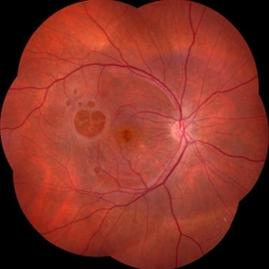
 Giant Persistent Macular Hole
Dec 6 2024 by César Adrián Gómez Valdivia, MD
Giant Persistent Macular Hole found in a 48 year-old male patient one year after vitrectomy.
Photographer: @eyemissu2
Imaging device: TOPCON TRC-50DX
Condition/keywords: macular, macular hole
-
 Traumatic Macular Hole pre and post repair
Nov 25 2024 by Shobhit Chawla, M.S.
31 year-old male reported with h/o of blunt trauma over right eye ,from cricket ball. On examination DVA RE 6/60,LE 6/18,ant segment BE :WNL,FUNDUS RE:Sub retinal hemorrhage at macula with chroidal tear,LE :WNL. Undwer went 25G vitrectomy+sub retinal TPA+C3F8(RE).Post op 1 month DVA RE:6/24 ,ANT SEGMENT:WNL,FUNDUS:resolved sub retinal haem with traumatic macular hole. Under went repeat vit+autologous retinal transplant +SOI RE.POST SOR AFTER4monthsV/A :6/18 RE
Photographer: Ranjit Ray
Imaging device: Clarus 500
Condition/keywords: Macular hole, retinal graft, subretinal hemorrhage
-
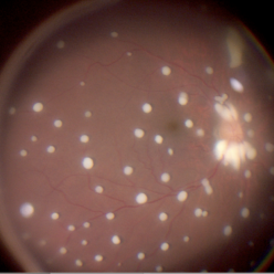
 Galaxy
Oct 29 2024 by SHILPI H NARNAWARE, ICO ( Retina) , FAICO ( Vitreo-Retina)
Retro mode image of a patient who underwent vitrectomy for RRD , with sub-retinal multiple PFCL bubbles giving appearance of arrangement of planets & other bodies in a galaxy
Photographer: Shilpi Narnaware, Sarakshi Netralaya , Nagpur, Maharashtra , India
Imaging device: Mirante ( by Nidek)
Condition/keywords: PFCL
-
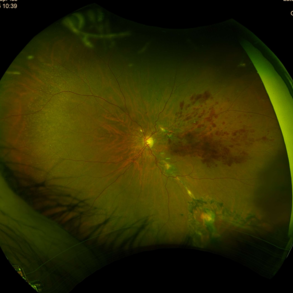
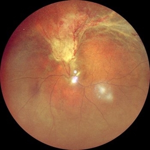
 Morning Glory Anomaly With Retinal Detachment Managed With Amniotic Membrane Graft
Oct 15 2024 by Hemanth Murthy, MBBS, MD, FASRS
10 year-old boy presented with noticed blurring of vision. He had total retinal detachment with complicated cataract. He underwent lensectomy with 240 band and vitrectomy with silicone oil. The retina failed to settle due to minute breaks in the inferior part of the disc. Repeat surgery with AMG was done to cover the inferior part of disc. The retina settled under silicone oil. Silicone oil was removed and he is presently undergoing amblyopia treatment. Vision is 2/60 with +14.5 diopter lens.
Photographer: Mr Veda Vyas
Condition/keywords: amniotic membrane graft, Morning Glory Anomaly
-
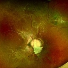
 Persistent Fetal Vasculature
Sep 23 2024 by Carlos Augusto Moreira, MD, PhD
Persistent Fetal Vasculature - an intraocular cotton swab appearance.
Photographer: Carlos Augusto Moreira-Neto, Hospital de Olhos do Paraná
Imaging device: NGENUITY Visualization System
Condition/keywords: pars plana vitrectomy (PPV), persistent fetal vasculature (PFV)
-
 Inverted ILM flap in RD with FTMH-First Post Op Day
Aug 23 2024 by SHILPI H NARNAWARE, ICO ( Retina) , FAICO ( Vitreo-Retina)
This is first post op day OCT of patient showing stuffed ILM in macular hole ,who underwent Vitrectomy with inverted ILM flap with silicon oil insertion in a case of RRD with FTMH.
Photographer: Shilpi Narnaware, Sarakshi Netralaya , Nagpur, Maharashtra , India
Imaging device: Mirante ( by Nidek)
Condition/keywords: FTMH, Inverted ILM technique, RD
-
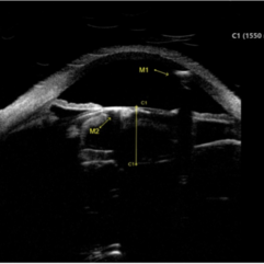 Intralenticular Gas
Aug 10 2024 by Varsha Reddy
Ultrasound biomicroscopy image of gas in the anterior chamber (A, M1) and lenticular cortex (A, M2) of a 68-year-old man following pneumatic retinopexy. Patient presented with a macula-off retinal detachment requiring vitrectomy, with pneumatic retinopexy done in office at post-operative week 1 to supplement poor gas fill.
Condition/keywords: intralenticular gas, pneumatic retinopexy, post-op
-
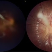
 RD with PVR in CMV Retinitis in an HIV Positive Patient
Jul 31 2024 by Tejaswita Verma
Fundus photograph of a 48 year old male with CF 1.5 mt vision having history of CMV retinitis, on HAART with CD4 count 81, showing retinal detachment with proliferative vitreoretinopathy changes. He was advised pars plana vitrectomy with silicon oil infusion.
Photographer: DR. TEJASWITA VERMA
Imaging device: MIRANTE
Condition/keywords: CMV retinitis with retinal detachment, HIV
-
 Posterior-PFV
Jul 27 2024 by Gokcen Deniz Gulpinar Ikiz
7 Year old girl presented with blurred vision on the left eye, with intermittent esotopia. She had been followed conservatively for intermittent esotropia on the left eye, recently advised for patching of the right eye. The vision is 1.0 on the right eye and 0.4 (Snellen) on the left eye. Anterior segment is natural bilaterally, except 20 PD esotropia on the left eye, with alternation and fixation. Refraction was +0.25 +0.25 x180 and +1.00-1.50 x60 on the right and left eyes respectively. Dilated fundus examination was natural on the right eye. However, there was a fibrotic stalk originating from the optic nerve head extending to the vitreous, terminating in the middle of the vitreous cavity, in a spider web configuration. Which also causes nasal dragging of the macula, leading to partial shallow detachment of the fovea nasally. Vitrectomy is advised for the left eye, with lens preserving approach, to preserve the current functional potential and the anatomy of the globe in long term.
Photographer: Gokcen Deniz Gulpinar Ikiz, Special Eye Clinic
Condition/keywords: amblyopia, posterior PFV, vitrectomy
-
 RPE-Transplantation
Jul 25 2024 by Gabriel Costa Andrade, PhD
Postoperative period of RPE-transplantation in a patient with neovascular AMD after RPE tear.
Photographer: Gabriel Andrade
Condition/keywords: neovascular age-related macular degeneration (AMD), pars plana vitrectomy (PPV), wet age-related macular degeneration (wet AMD)
-
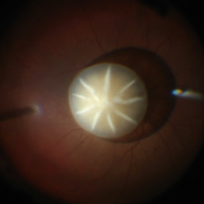
 Failure of Macular Hole Surgery
Jul 2 2024 by Abel Ramírez-Estudillo, MD
Fundus photograph of a 67-year-old woman with failed macular hole surgery, now referred to our clinic with 8 holes.
Photographer: Berenice Palafox, Centro Oftalmológico Mira, Mexico City
Imaging device: Zeiss
Condition/keywords: iatrogenic retinal tear, internal limiting membrane (ILM) peeling, macular hole, vitrectomy
-
 Post-op Vitreous Surgery
Jun 22 2024 by Sanauddin Samejo , Diploma (Ophthalmic Technician Training Course)
A 51 years Old Male visited after Vitreous Surgery.
Photographer: Sanauddin Samejo, Burjeel Hospital, Abu Dhabi, UAE
Imaging device: Optos Silver Stone
Condition/keywords: post-op, vitrectomy
 Loading…