 Initializing download.
-
 By Rita Couceiro, MD, MS
Co-author(s): Carlos Marques Neves, MD PhD, Hospital de Santa Maria, Lisboa, Portugal, Joaquim Prates Canelas, MD, Hospital de Santa Maria, Lisboa, Portugal - Uploaded on Mar 23, 2015.
- Last modified by Caroline Bozell on Aug 7, 2015.
- Image of the week
-
Aug 9, 2015
View all images of the week - Rating
- Appears in
- Miscellaneous
- Condition/keywords
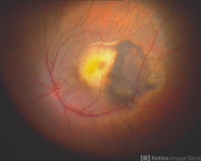
- submacular hemorrhage, wet age-related macular degeneration (wet AMD), neovascular age-related macular degeneration (AMD)
- Imaging device
-
Fundus camera
Intraoperative fundus photograph - Description
- An 80-year-old male, with a history of hypertension and high cholesterol, complained of acute and painless vision loss in his left eye (OS) in the previous 5 months. On observation best corrected visual acuity in OS was hand motion. A dense vitreous opacity in OS precluded fundus examination. Ocular ultrasound revealed vitreous hemorrhage and thickening of the macular area. The patient was submitted to pars plana vitrectomy, which disclosed a large submacular hemorrhage with chronical features and disciform scarring in the setting of neovascular AMD.


