Search results (26 results)
-
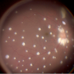 Seedlings of Fungal Endophthalmitis
Mar 14 2025 by SHILPI H NARNAWARE, ICO ( Retina) , FAICO ( Vitreo-Retina)
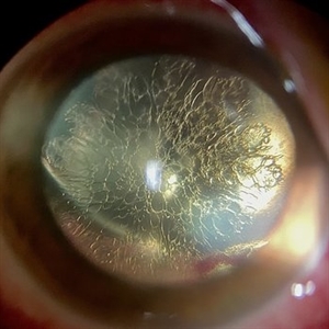
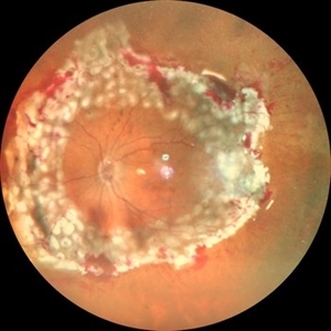
57 year diabetic female , was treated as a case of recurrent vitreous post cataract surgery. Patient was posted for vitrectomy 3 months post cataract surgery. Intra-operatively, multiple yellowish colonies were seen all over the posterior pole were seen, which were later found to be Aspergillus colonies.
Photographer: Shilpi Narnaware, Sarakshi Netralaya , Nagpur, Maharashtra , India
Imaging device: Ngenuity
Condition/keywords: endophthalmitis, fungal
-
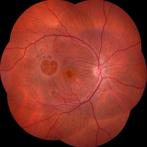 Failure of Macular Hole Surgery
Jul 2 2024 by Abel Ramírez-Estudillo, MD
Fundus photograph of a 67-year-old woman with failed macular hole surgery, now referred to our clinic with 8 holes.
Photographer: Berenice Palafox, Centro Oftalmológico Mira, Mexico City
Imaging device: Zeiss
Condition/keywords: iatrogenic retinal tear, internal limiting membrane (ILM) peeling, macular hole, vitrectomy
-
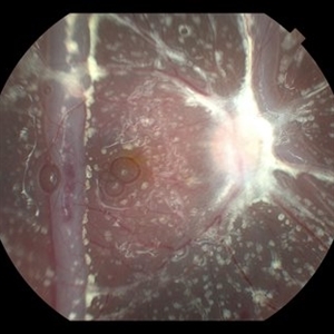 Fish Hook Eye Trauma
Jun 12 2024 by Miguel Brito, MD, FASRS
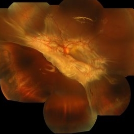
Fundus photograph of a 15-year-old boy post cataract aspiration, pars plana vitrectomy, suprachoroidal drainage, and retinal reattachment surgery secondary to traumatic endophthalmitis.
Photographer: Miguel Brito
Condition/keywords: endophthalmitis, PFCL, Retinal detachment under Silicon Oil, retinal fold
-
 Dislocated Lens, Posterior OD
Jan 26 2024 by Corey Grant
OPTOS California photo presents a 71 year old male patient with a dislocated lens, posterior in the right eye. Presented on 1/26/24 with posteriorly dislocated SN60WF with a Soemmerring ring. Associated retinal hemorrhage within retinoschisis as well. This will result in a PPV/IOL exchange/SFIOL/STK for the right eye.
Photographer: Corey Grant, Ophthalmic Imager, Retina Specialist of Michigan
Imaging device: OPTOS California
Condition/keywords: color photo, IOL, OD, Optos, OPTOS CALIFORNIA, pars plana vitrectomy (PPV), retina
-
 Retinal Fold
Sep 26 2023 by Mauricio Bayram-Suverza, MD
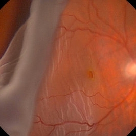
A 38-year-old man underwent vitrectomy in the left eye due to a giant tear in the upper retina. SF6 gas was used as endotamponade. During the post-surgical check-up, it was identified that the patient developed a full-thickness retinal fold due to retinal slippage during fluid-air exchange. As the fold was away from the macular area, it was decided to observe the patient. Three weeks after the surgery, his best-corrected visual acuity was 20/30.
Photographer: Mauricio Bayram-Suverza, Fundación Hospital Nuestra Señora de la Luz
Imaging device: TRC-50DX
Condition/keywords: giant retinal tear, retina surgery complications, Retinal slippage, vitreoretinal surgery
-
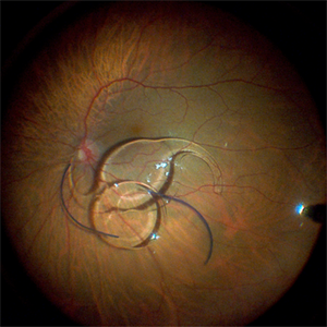 Fraternal Twins
May 22 2023 by Gustavo M. Hüning, MD, MBA, FASRS
Intrasurgical photograph using a non-contact system and 3D visualization system of a 65-year-old woman who suffered an ocular trauma.
Photographer: Gustavo M. Hüning, Hüning Clínica do Olhar, Santa Maria - Brazil
Imaging device: Alcon Luxor combined with Alcon nGenuity
Condition/keywords: dislocated intraocular lens (IOL), implant, pars plana vitrectomy (PPV)
-
 Feather like cataract
Apr 11 2023 by rodrigo torres
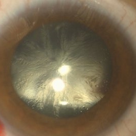
Cataract after vitrectomy and gas tamponade.
Photographer: Rodrigo Amaral Torres
Condition/keywords: cataract, pars plana vitrectomy (PPV)
-
 Dislocated Lens
Apr 26 2023 by Chloe Hanifan
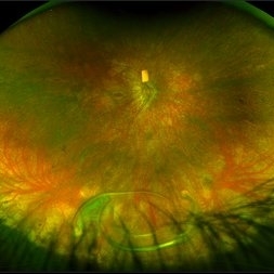
Ultra wide field fundus photograph of a 41-year-old male with a dislocated lens affecting his right eye. IOL noted inferior vitreous base and vitrectomy surgery for removal of IOL was recommended. Patient has history of retinitis pigmentosa as well. Patient's vision at the time of presentation was counting fingers at 2 feet.
Photographer: Chloe Hanifan
Imaging device: Optos California
Condition/keywords: dislocated lens, fundus photography, Optos, pseudocolor, retinitis pigmentosa, ULTRA WIDE FIELD
-
 Lady in a dress
Feb 9 2023 by Shelby Helton
Fluorescein Angiography on a 67-year-old male with significant RPE changes secondary to a severe subretinal hemorrhage that required a vitrectomy with subretinal TPA in 2013.
Photographer: Shelby Helton
Imaging device: Heidelberg Spectralis
Condition/keywords: wet age-related macular degeneration (wet AMD)
-
 High risk Proliferative Diabetic Retinopathy treated with Pan Retinal Photocoagulation
Nov 5 2022 by Somnath Chakraborty, MD
A Fundus Photo Montage of 43 year old Asian Male with Type 2 Diabetes Mellitus since 7 years who presented with sudden onset diminition of vision in his Left eye. BCVA OS was 20/200. He was diagnosed to have Pre retinal bleed due to Proliferative Diabetic Retinopathy and was treated with Pan Retinal Photocoagulation. This image shows a large neo-cascular frond at the disc and superior to it with Pre-retinal bleed and Fresh laser marks along
Photographer: Pulak Roy
Condition/keywords: diabetic blindness, diabetic retinopathy vitrectomy study (DRVS), fresh laser burns, laser photocoagulation, preretinal hemorrhage, proliferative diabetic retinopathy (PDR)
-
 submacular perfluorocarbon liquid
Sep 7 2022 by JEFFERSON R SOUSA, Tecg.º (Biomedical Systems Technology)
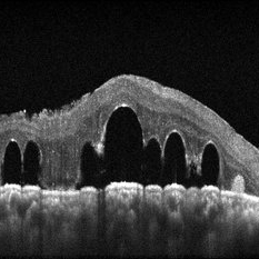
A 63-year-old male patient underwent vitreoretinal surgery with the use of perfluorocarbon. From a technological point of view, extended-field retinography presents many points of focus variation due to the difficulty of establishing a diffuse focus, as it is a recent post-operative case. In OCT Fundus Enface, although it has a low resolution, it is extremely important for documenting the presence of perfluor. Best seen in structural OCT.
Photographer: JEFFERSON ROCHA DE SOUSA - Retinal Department at Instituto Dr. Suel Abujamra Sao Paulo-Brazil
Imaging device: Optical Coherence Tomography system OCT CIRRUS 5000, Protocol, HD 5 Line
Condition/keywords: perfluorocarbon fluid, post-vitrectomy, submacular perfluorocarbon liquid (PFO), vitrectomy
-
 Subretinal Bleed
Jul 12 2022 by Akansha Sharma
73 year old diabetic and hypertensive female presented with sub-retinal hemorrhage for which she was operated with pars-plana vitrectomy with intra-vitreal anti-VEGF
Photographer: Dr. Akansha Sharma-Retina Foundation, Ahmedabad
Condition/keywords: subretinal hemorrhage, subretinal blood
-
 JXT and Proliferative Diabetic Retinopathy
Jan 13 2022 by ASRS Staff
Wide field photograph of 50 year-old woman, known case of JXT in both eyes and known diabetic, after 9 months of PPV for subhyaloid hemorrhage.
Imaging device: Nidek Mirante
Condition/keywords: florid type PDR, JXT, pars plana vitrectomy (PPV)
-
 360 Degree Retinectomy
Sep 11 2020 by Sham Talati, DOMS
A case of retinal detachment with PVR. Patient underwent pars plana vitrectomy with silicon oil injection with 360 degree retinectomy.
Photographer: Dr. Sham Talati,Retina Foundation,Ahmedabad
Imaging device: Nidek Mirante
Condition/keywords: proliferative vitreoretinopathy (PVR), retinectomy
-
 Diabetic Macular TRD
Jan 10 2020 by Somnath Chakraborty, MD
Fundus Montage image of the left eye of a 48-year-old type 2 diabetic with post PRP macular extensive tractional retinal detachment involving macula.
Photographer: Pulak Roy
Condition/keywords: diabetic retinopathy, proliferative diabetic retinopathy (PDR), tractional retinal detachment, vitrectomy, vitreomacular surgery
-
 Retinal Detachment with Giant Retinal Tear and Macular Hole
Jan 6 2020 by MATTEO FORLINI, MD
A 61-year-old-male patient presented with sudden diminution of vision in the right eye due to retinal detachment with giant retinal tear and macular hole. Best corrected visual acuity (BCVA) at presentation was 20/200. A 23 G vitrectomy was performed. The edges of the tear were unrolled and complete retinal re-attachment under PFCL was achieved. A 360 degree intraoperative endolaser was performed on the peripheral retina as well as around the edges of the tears. PFCL was exchanged with silicone oil 5000cs as final tamponade. At six-months follow-up retina was attached and macular hole was repaired. Best-corrected visual acuity is 20/125 at present.
Photographer: Matteo Forlini MD, San Marino Hospital, Republic of San Marino
Condition/keywords: full thickness macular hole, giant retinal tear, silicone oil
-
 Vitreous Amyloidosis Slit Lamp Photo
Oct 23 2019 by Alexander D Port, MD
Slit lamp photograph preoperatively demonstrating dense symptomatic vitreous opacity in the setting of amyloidosis. The patient elected to undergo pars plana vitrectomy.
Condition/keywords: slit lamp photo, vitreous amyloidosis
-
 Submacular Hemorrhage
Apr 24 2018 by Pauline T Merrill, MD, FASRS
Fundus photo of left eye of a 65-year-old AMD patient who presented with sudden drop of vision from 20/30 to CF due to a large submacular hemorrhage, 7 months following her last Eylea injection. She underwent immediate injection of C3F8 in the office, with little effect. 10 days later vitrectomy with subretinal tPA and air-fluid exchange was performed, with successful displacement of the hemorrhage.
Photographer: Ermelinda Diaz, Illinois Retina Associates, Chicago, Illinois
Imaging device: Topcon 50DX
Condition/keywords: neovascular age-related macular degeneration (AMD), submacular hemorrhage
-
 Vasoproliferative Tumor With Resultant Total Exudative RD Status Post Vitrectomy/Laser/Oil/Dexamethasone Intravitreal Implant
Apr 25 2017 by Christopher G Fuller, MD
Fundus photograph of a presumptive vasoproliferative tumor (with resultant total exudative retinal detachment) in a 54-year-old white truck driver. Image is taken on post-operative day 4, after 25/27 gauge vitrectomy with drainage retinotomy, air-fluid exchange, endoscopic laser blanching of VPT, oil, and dexamethasone intravitreal implant.
Photographer: Ray Gardner, Texas Retina Associates (Lubbock, TX)
Condition/keywords: exudative retinal detachment
-
 Lens Feathering
Nov 9 2016 by Nimrod Dar
26-year-old man with lens feathering a day after a pars plana vitrectomy for rhegmatogenous retinal detachment.
Photographer: Nimrod Dar, M.D, Meir Ophthalmology Department
Condition/keywords: lens feathering, vitrectomy
-
 Retinal Detachment Repair With Silicone Oil and Scleral Buckle, Fourteen Years Later, With Visual Acuity of 20/25
Sep 12 2016 by Timothy S Fuller, MD
65-year-old woman s/p scleral buckle 14 years ago. Two weeks later, the retina re-detached, and vitrectomy, laser, and silicone oil procedure was performed. Patient remains 20/25 with correction fourteen years later. The cornea is clear, there is no oil emulsification, and there is a stable, moderately inferiorly subluxated PCIOL (as it was prior to RD surgery). IOP is 17 on Cosopt BID.
Photographer: Nicholas Hesse, Texas Retina Associates
Imaging device: Optos
Condition/keywords: laser, scleral buckle, silicone oil
-
 Proliferative Vitreoretinopathy Under Silicone Oil
Mar 15 2016 by Sjakon G Tahija, MD
This young patient developed PVR under silicone oil after traumatic vitrectomy for long standing retinal detachment.
Photographer: Avris Siahaan, Klinik Mata Nusantara
Condition/keywords: proliferative vitreoretinopathy (PVR), silicone oil
-
 IOFB Combined
Mar 12 2015 by Ahmad B. Tarabishy, MD
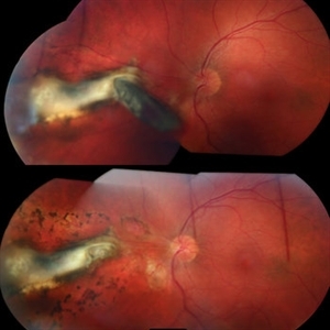
A 26-year-old gentleman presented with a metallic intraocular foreign body embedded in the nasal retina (above). Post-operative appearance two weeks after vitrectomy, foreign body removal, endolaser, and gas (below).
Photographer: Jessica Armbruster
Imaging device: Topcon TRC-50EX
Condition/keywords: encapsulated intraocular foreign body, non metallic retained intraocular foreign body (RIOFB), penetrating trauma
-
 Chronical Submacular Hemorrhage in the Setting of Neovascular AMD
Mar 23 2015 by Rita Couceiro, MD, MS
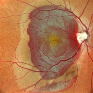
An 80-year-old male, with a history of hypertension and high cholesterol, complained of acute and painless vision loss in his left eye (OS) in the previous 5 months. On observation best corrected visual acuity in OS was hand motion. A dense vitreous opacity in OS precluded fundus examination. Ocular ultrasound revealed vitreous hemorrhage and thickening of the macular area. The patient was submitted to pars plana vitrectomy, which disclosed a large submacular hemorrhage with chronical features and disciform scarring in the setting of neovascular AMD.
Imaging device: Intraoperative fundus photograph
Condition/keywords: neovascular age-related macular degeneration (AMD), submacular hemorrhage, wet age-related macular degeneration (wet AMD)
-
 Optic Atrophy and Attenuated Retinal Vessels Following Endophthalmitis
Jul 12 2014 by Philip J. Polkinghorne, MD
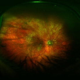
This elderly lady underwent a vitrectomy for post-surgical endophthalmitis. The infection was successfully treated but the functional outcome was poor because of optic atrophy and attenuated retinal vessels.
Photographer: Alex Fraser
Imaging device: Optos Camera
Condition/keywords: attenuated vessels, endophthalmitis, optic atrophy, post-vitrectomy
 Loading…