Search results (561 results)
-
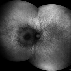
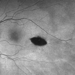 Torpedo Maculopathy
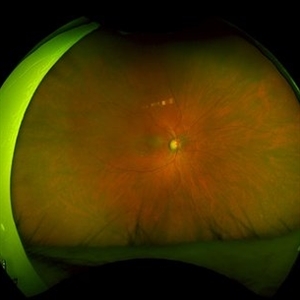
Feb 20 2024 by Soobien Lee
Optos fundus autofluorescence photograph of a 35-year-old asymptomatic female with no ocular or medical history with stable and chronic appearing torpedo-shaped macula lesion in the left eye.
Photographer: Peter Sotirakos, Elman Retina Group
Imaging device: Optos Ultra-Widefield Autoflurescence Imaging
Condition/keywords: autofluorescence imaging, genetics, macula, maculopathy, Optos, torpedo maculopathy
-
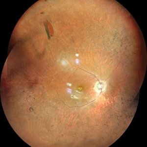
 Torpedo Maculopathy
Feb 20 2024 by Soobien Lee
Optos color fundus photograph of a 35-year-old asymptomatic female with no ocular or medical history with stable and chronic appearing torpedo-shaped macula lesion in the left eye.
Photographer: Peter Sotirakos, Elman Retina Group
Imaging device: Optos Ultra-Widefield Imaging
Condition/keywords: macula, Optos, torpedo maculopathy
-
 "The Eye of Sauron"
Mar 14 2023 by Anfisa Ayalon, MD
Fundus autofluorescence image of a 38-year-old female with “Bull's eye” pattern maculopathy. There is no history of medication use associated with retinal toxicity. BCVA RE 20/25+2
Photographer: Danielle Ferguson and Alec Bertoni, University of Pittsburgh Medical Center
Condition/keywords: bull's eye maculopathy, retina
-
 Hypotony Maculopathy
Nov 3 2023 by Matthew Dombrow, MD
31 year old female 4 days s/p Ahmed Valve
Photographer: Cori Sturtevant, Connecticut Retina Consultants, Hamden, Connecticut
Imaging device: Optos - California
Condition/keywords: hypotony maculopathy
-
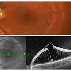
 Myopic Traction Maculopathy
Mar 17 2025 by Drew Mitchell
HD 1 line 100x 9 mm scan of a right eye with MTM at stage 3c. Macular Schisis Detachment.
Photographer: Drew Mitchell OCT-C
Imaging device: Zeiss Cirrus 5000
Condition/keywords: full thickness macular hole, Macular hole, myopic foveoschisis, myopic macular schisis, myopic traction maculopathy, PVD
-
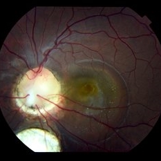
 Optic Disc Pit with Maculopathy
Feb 25 2021 by Niloofar Piri, MD
Color fundus photograph and SD OCT of a 6-year-old patient with optic disc pit associated with large retinoschisis involving the entire macula. SD OCT demonstrating large retinoschisis with ILM draping centrally giving it the appearance of pseudohole on the corresponding central area of color photo. Vision 20/80
Photographer: Lisa Breeding, St Louis University
Condition/keywords: maculopathy, optic disc
-
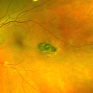
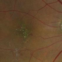 West African Crystalline Maculopathy
Oct 22 2023 by Niloofar Piri, MD
Fundus photograph of right eye of a patient from Liberia demonstrating multiple birefringent yellow green crystalline deposits in the fovea. Please notice the partially fibrosed NVE inferiorly. The disease has been shown to be associated with vascular disorders including diabetic retinopathy.
Photographer: Niloofar Piri, MD
Condition/keywords: crystalline maculopathy, crystalline retinopathy, West African Crystalline maculopathy
-
 Advanced PDR RE FFA
Aug 31 2014 by Neha Goel, MS DNB FRCS (Glasg)
Fluorescein angiogram of the right eye.
Photographer: Neha Goel
Imaging device: Zeiss Visucam
Condition/keywords: fibrovascular proliferation, ischaemic diabetic maculopathy, proliferative diabetic retinopathy (PDR)
-
 Advanced PDR-RE
Aug 31 2014 by Neha Goel, MS DNB FRCS (Glasg)
Fundus photograph of the right eye of a 50-year-old diabetic male.
Photographer: Neha Goel
Imaging device: Zeiss Visucam
Condition/keywords: fibrovascular proliferation, ischaemic diabetic maculopathy, proliferative diabetic retinopathy (PDR)
-
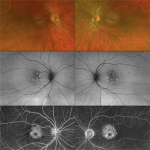
 Bilateral “Bull's eye”pattern maculopathy
Mar 14 2023 by Anfisa Ayalon, MD
Both eyes fundus autofluorescence image of a 38-year-old female with “Bull's eye” pattern maculopathy. There is no history of medication use associated with retinal toxicity. BCVA RE 20/25+2, LE 20/20-3
Photographer: Danielle Ferguson and Alec Bertoni, University of Pittsburgh Medical Center
Condition/keywords: bull's eye maculopathy, maculopathy, retina
-
 Bilateral Benign Yellow Dot Maculopathy
May 6 2025 by Amol yuvraj ganvir
A 37-year-old female patient presented for a routine eye examination. Her best-corrected visual acuity was 6/6 in both eyes. Fundus examination revealed multiple small yellow dots over the macula in both eyes. FAF imaging demonstrated characteristic hyperautofluorescence corresponding to these dots.
Photographer: Dr. Amol Ganvir, Vitreo-Retina Fellow, Ishwar Eye Centre, Rohtak, Haryana
Imaging device: Visucam-Zeiss
Condition/keywords: Autoflourescence, yellow dots
-
 Bulls Eye
Dec 2 2012 by Rogerio N Shinsato, MD, PhD
central areolar choroidal dystrophy, bulls eye
Photographer: rogerio neri shinsato
Condition/keywords: bull's eye maculopathy, central areolar choroidal dystrophy (CACD)
-
 CERKL-related Cone Rod Dystrophy
Jun 27 2022 by Hanna Choi
37-year-old female with cone-rod dystrophy. Developed photophobia and progressive blurry vision in the third decade. VA 20/40 OD, 20/30 OS. The patient is compound heterozygous for pathogenic mutations in the CERKL gene (Arg465Trp and Arg283*).
Photographer: Kaitlynn Silva, New England Retina Consultants
Imaging device: Ultrawide-field Optos Fundus Photography, Autofluorescence, Fluorescein Angiography
Condition/keywords: cone dystrophy, inherited retinal disease, maculopathy
-
 Chloroquine maculopathy
Jun 22 2022 by JORGE SOBERANES
Fundus autofluorescence of a bull´s eye maculopathy of a 55-year-old woman treated for ten years with choloquine.
Photographer: Jorge I. Soberanes MD, Asociación para Evitar la Ceguera en México.
Imaging device: Zeiss Clarus 700 (Green autofluorescence)
Condition/keywords: bull's eye maculopathy, chloroquine, fundus autofluorescence (FAF), macula, maculopathy
-
 Chloroquine maculopathy 1
Jan 11 2013 by Alex P. Hunyor, MD
Chloroquine maculopathy 1 - color image, right eye.
Condition/keywords: chloroquine maculopathy, chloroquine toxicity
-
 Chloroquine maculopathy 5
Jan 11 2013 by Alex P. Hunyor, MD
Chloroquine maculopathy 5 - fluorescein angiogram, left eye.
Condition/keywords: chloroquine maculopathy, chloroquine toxicity
-
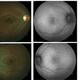
 Colobomatous Optic Disc Maculopathy
Feb 13 2020 by Yoshihiro Yonekawa, MD, FASRS
Beautifully focused fundus photograph of a teenage girl with submacular fluid from a colobomatous optic disc.
Photographer: Netanya Lerner, COA, Wills Eye Hospital/Mid Atlantic Retina
Imaging device: Topcon
Condition/keywords: chorioretinal coloboma, coloboma of optic disc, congenital optic nerve pit, subretinal fluid
-
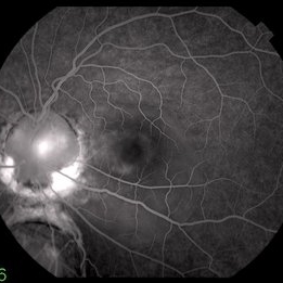 Colobomatous Optic Disc Maculopathy
Feb 13 2020 by Yoshihiro Yonekawa, MD, FASRS
Fluorescein angiography, late frame, of a teenage girl with submacular fluid from a colobomatous optic disc. The camera is focused is on the elevated macula, and the disc is subtly defocused.
Photographer: Netanya Lerner, COA, Wills Eye Hospital/Mid Atlantic Retina
Imaging device: Topcon
Condition/keywords: chorioretinal coloboma, coloboma of optic disc, congenital optic nerve pit, subretinal fluid
-
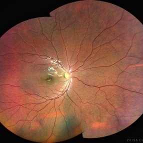 Cone Dystrophy
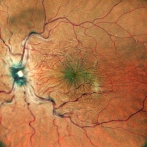
Aug 30 2023 by Vishal Agrawal, MD, FRCS,FACS,FASRS
12 year old male patient presented with photophobia, decrease in vision and Nystagmus. Bulls eye maculopathy gives an appearance of an eye on the fovea on color fundus photo due to nystagmus.
Photographer: Dr Bhagyashree
Imaging device: Clarus 700
Condition/keywords: bull's eye maculopathy, Cone-Rod Dystrophy
-
 Focal Retinitis- Unilateral Acute Idiopathic Maculopathy OS.
Nov 13 2019 by Tammy Mclaughlin
No prodromal URI. Symptoms present for 1 1/2 weeks. No serous detachment presently. Does have typical circular RPE changes in macula without leakage or edema. Typical course is spontaneous improvement with residual RPE changes. Recommended observation.
Photographer: Tammy Mclaughlin
Imaging device: Zeiss Visucam
Condition/keywords: focal retinitis, unilateral acute idiopathic maculopathy
-
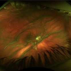
 Horseshoe Tear in Retinitis Pigmentosa
Mar 22 2021 by ASRS Staff
Montage of 25-year-old patient, high myopic patient came with complaint of diminution of vision in both eyes and on posterior segment examination of right eye, HST was present along with maculopathy.
Imaging device: Nidek Mirante
Condition/keywords: maculopathy, retinitis pigmentosa
-
 Hydroxychloroquine Maculopathy
Jul 23 2023 by Ahmad B. Tarabishy, MD
62 year old female with rheumatoid arthritis, treated with hydroxychloroquine 200 mg BID for the past 6-8 years. She presents with blurred vision, difficulty reading, and difficulty transitions from dark to light conditions since 4 months.
Photographer: Dr. Angela Rico
Condition/keywords: hydroxychloroquine toxicity, plaquenil toxicity, toxic maculopathy
-
 Hypotonous Maculopathy
Jul 12 2022 by Akansha Sharma
FUNDUS PHOTOGRAPH OF A 37 YEAR OLD MALE WITH HISTORY OF BLUNT TRAUMA WITH TENNIS BALL PRESENTING WITH HYPOTONOUS MACULOPATHY
Photographer: Dr. Akansha Sharma-Retina Foundation, Ahmedabad
Condition/keywords: hypotonous retinopathy
-
 Laser Pointer Maculopathy
Jul 1 2018 by John S. King, MD
9-year-old with two month history of decreased vision that has improved some over time. 20/20 OD and 20/30 OS. Further prompting, he admitted to playing with "toy lasers" with his friends recently. He also looked at the eclipse, but used protective eye wear. Photos show some RPE defects characterized by a hyperpigmented center with hypopigmented halo.
Photographer: Karin Aletter
Imaging device: Topcon
Condition/keywords: laser pointer maculopathy, laser pointer retinopathy, maculopathy
-
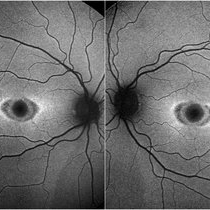
 PAMM-OCTA
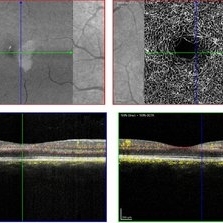
Nov 29 2023 by Daniel Davis, OCT-C
OCT-A of a 30 yo female with PAMM OD.
Photographer: Daniel Davis, OCT-C
Imaging device: Heidelberg Spectralis
Condition/keywords: OCTA, paracentral acute middle maculopathy
 Loading…