 Initializing download.
-
 By Niloofar Piri, MD
SSM Health Group, St Louis University
Co-author(s): Katherine Bussan, MD, St Louis University - Uploaded on Feb 25, 2021.
- Last modified by Caroline Bozell on Feb 26, 2021.
- Rating
- Appears in
- Miscellaneous
- Condition/keywords
- optic disc, maculopathy
- Photographer
- Lisa Breeding, St Louis University
- Description
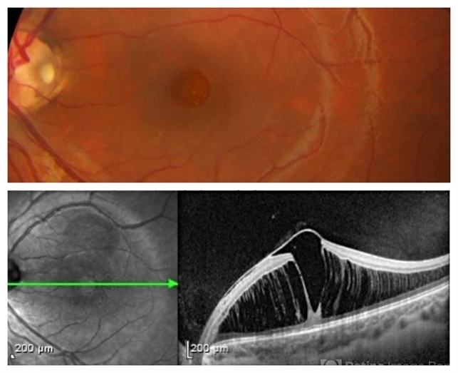
- Color fundus photograph and SD OCT of a 6-year-old patient with optic disc pit associated with large retinoschisis involving the entire macula. SD OCT demonstrating large retinoschisis with ILM draping centrally giving it the appearance of pseudohole on the corresponding central area of color photo. Vision 20/80

---thumb.jpg/image-square;max$79,0.ImageHandler "Melanocytoma")
---thumb.jpg/image-square;max$79,0.ImageHandler "Optic Disc and Retinal Edema")


---thumb.jpg/image-square;max$79,0.ImageHandler "Optic Nerve Mielinization")


