Search results (561 results)
-
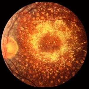
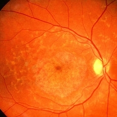 Fundus Flavimaculatus
May 2 2013 by Henry J. Kaplan, MD
Yellow pisciform flecks at the level of RPE accompanied by Bull's eye, Right eye; #1.
Condition/keywords: bull's eye maculopathy, fundus flavimaculatus, Stargardt disease
-
---thumb.jpg/image-square;max$300,300.ImageHandler) Bull's Eye Maculopathy
Bull's Eye Maculopathy
Feb 20 2013 by From the Collections of Thomas M. Aaberg, MD and Thomas M. Aaberg Jr., MD
bull's eye maculopathy fundus photo
Condition/keywords: bull's eye maculopathy
-
---thumb.jpg/image-square;max$300,300.ImageHandler) Tamoxifen Retinopathy- OCT
Tamoxifen Retinopathy- OCT
Aug 30 2012 by Young Hee Yoon, MD, PhD
OCT image of an 58-year-old woman with a bilateral tamoxifen maculopathy. She had taken tamoxifen for 24 months due to breast cancer. In spite of discontinuation 2 years ago, her macula remained unchanged. Her best-corrected visual acuity was 20/50 in the right and 20/100 in the left.
Photographer: Soon Tae Kim, Asan Medical Center
Imaging device: Heidelberg Spectralis
Condition/keywords: drug toxicity
-
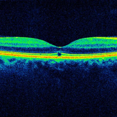 Solar Maculopathy, OCT, Right Macula
Mar 7 2015 by Thomas A. Ciulla, MD, MBA, FASRS
OCT revealed symmetric focal discontinuity of the IS/OS line and underlying RPE.
Condition/keywords: solar maculopathy, solar retinopathy
-
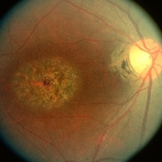 Stargardt's Disease
May 2 2013 by Henry J. Kaplan, MD
Fundus photograph of the right eye in a patient with Stargardt's disease shows typical bull's eye maculopathy; #1.
Condition/keywords: bull's eye maculopathy, Stargardt disease
-
 OCT Myopic Staphyloma With Schisis and ERM
Apr 24 2014 by Scott E. Pautler, MD
OCT of high myope with asymptomatic macular schisis.
Imaging device: Heidelberg Spectralis
Condition/keywords: foveal schisis, maculopathy, maculoschisis, optical coherence tomography (OCT), pathologic myopia, staphyloma
-
 Tamoxifen Retinopathy- OCT
Aug 30 2012 by Young Hee Yoon, MD, PhD
OCT image of an 58-year-old woman with a bilateral tamoxifen maculopathy. She had taken tamoxifen for 24 months due to breast cancer. In spite of discontinuation 2 years ago, her macula remained unchanged. Her best-corrected visual acuity was 20/50 in the right and 20/100 in the left.
Photographer: Soon Tae Kim, Asan Medical Center
Imaging device: Zeiss cirrus HD-OCT 4000
Condition/keywords: drug toxicity, toxic maculopathy
-
 Stargardt's disease
May 2 2013 by Henry J. Kaplan, MD
The same patient , left eye #2
Condition/keywords: bull's eye maculopathy, Stargardt disease
-
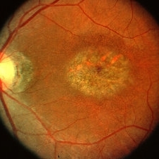
 Cone Dystrophy
Mar 29 2013 by Henry J. Kaplan, MD
Fundus photograph of a patient with low vision and hemeralopia and typical bull`s eye in cone dystrophy #2.
Condition/keywords: bull's eye maculopathy, cone dystrophy
-
---thumb.jpg/image-square;max$300,300.ImageHandler) Tamoxifen Retinopathy- Fundus photo
Tamoxifen Retinopathy- Fundus photo
Aug 30 2012 by Young Hee Yoon, MD, PhD
Fundus photograph of an 58-year-old woman with a bilateral tamoxifen maculopathy. She had taken tamoxifen for 24 months due to breast cancer. In spite of discontinuation 2 years ago, her macula remained unchanged. Her best-corrected visual acuity was 20/50 in the right and 20/100 in the left.
Photographer: Sung Hyun Kim, Asan Medical Center
Imaging device: Topcon
Condition/keywords: drug toxicity, toxic maculopathy
-
 Stargardt's Disease
Oct 8 2012 by Susanna S. Park, MD, PhD
Fluorescein angiogram of a 12-year-old girl with progressive loss of vision in both eyes.
Photographer: Ellen Redenbo, University of California Davis Eye Center
Condition/keywords: bull's eye maculopathy, dark area on retina and choroid, Stargardt disease
-
 Cone Dystrophy
Mar 29 2013 by Henry J. Kaplan, MD
Fundus photograph of a patient with progressive vision loss and hemeralopia (cone dystrophy) shows bull`s eye pattern #1.
Condition/keywords: bull's eye maculopathy, cone dystrophy
-
 Stargardt's Disease
Oct 8 2012 by Susanna S. Park, MD, PhD
Fundus photograph of a 12-year-old girl with progressive loss of vision in both eyes.
Photographer: Ellen Redenbo, University of California Davis Eye Center
Condition/keywords: bull's eye maculopathy, dark area on retina and choroid, Stargardt disease
-
 Tamoxifen Maculopathy
-
 Tamoxifen Retinopathy- FAF
Aug 30 2012 by Young Hee Yoon, MD, PhD
Fundus autofluorescence (FAF) of an 58-year-old woman with a bilateral tamoxifen maculopathy. She had taken tamoxifen for 24 months due to breast cancer. In spite of discontinuation 2 years ago, her macula remained unchanged. Her best-corrected visual acuity was 20/50 in the right and 20/100 in the left.
Photographer: Soo Hyun Cho, Asan Medical Center
Imaging device: Heidelberg HRA II
Condition/keywords: drug toxicity, toxic maculopathy
-
 Plaquenil Toxicity
Apr 30 2013 by Theodore Leng, MD, MS, FASRS
Color fundus photograph from a 44-year-old woman with bilateral plaquenil toxicity. A mild bulls-eye maculopathy is present.
Condition/keywords: hydroxychloroquine toxicity, plaquenil toxicity
-
 Tamoxifen Retinopathy- Fundus photo
Aug 30 2012 by Young Hee Yoon, MD, PhD
Fundus photograph of an 58-year-old woman with a bilateral tamoxifen maculopathy. She had taken tamoxifen for 24 months due to breast cancer. In spite of discontinuation 2 years ago, her macula remained unchanged. Her best-corrected visual acuity was 20/50 in the right and 20/100 in the left.
Photographer: Ji Hee Kim, Asan Medical Center
Imaging device: Canon CR-DGI
Condition/keywords: drug toxicity, toxic maculopathy
-
 Bulls eye retinopathy
Nov 20 2012 by Roy Schwartz, MD
75-YEAR-OLD FEMALE PRESENTS WITH BILATERAL GRADUAL VISUAL LOSS 6/30 Dx BE PSEUDOPHAKIA + PCO BE BULLS EYE MACULOPATHY PER FA VA IMPROVES TO 6/10 S/P YAG CAPSULOTOMY OCT - BE MACULAR SUBRETINAL FLUID NO HISTORY OF CHLOROQUINE THERAPY NO DRUSEN OR SIGNS OF AMD WORKING DIAGNOSIS - BE CHRONIC CSCR
Imaging device: Heidelberg spectralis
Condition/keywords: bull's eye maculopathy, optical coherence tomography (OCT)
-
 Torpedo Maculopathy 1
Feb 26 2014 by Raj K. Maturi, MD
Color fundus of a 47-year-old female with torpedo maculopathy- atypical choroidal nevus.
Photographer: Charlotte Harris COA Midwest Eye Institute Indianapolis, Indiana
Imaging device: TOPCON 50EX
Condition/keywords: choroidal nevus, fundus photograph, torpedo maculopathy
-
 Bulls Eye
Dec 2 2012 by Rogerio N Shinsato, MD, PhD
central areolar choroidal dystrophy, bulls eye
Photographer: rogerio neri shinsato
Condition/keywords: bull's eye maculopathy, central areolar choroidal dystrophy (CACD)
-
 Torpedo maculopathy
Jan 11 2013 by Alex P. Hunyor, MD
Torpedo maculopathy, left eye.
Condition/keywords: torpedo maculopathy
-
 Plaquenil Toxicity
Apr 30 2013 by Theodore Leng, MD, MS, FASRS
Color fundus photograph from a 44-year-old woman with bilateral plaquenil toxicity. A mild bulls-eye maculopathy is present.
Condition/keywords: hydroxychloroquine toxicity, plaquenil toxicity
-
---thumb.jpg/image-square;max$300,300.ImageHandler) Primary Hyperoxaluria and Oxalosis
Primary Hyperoxaluria and Oxalosis
Jul 24 2013 by Hamid Ahmadieh, MD
Color fundus photograph of the right eye of a 55-year-old man with primary hyperoxaluria and oxalosis. Characteristic crystalline retinopathy (flecked retina), black geographic maculopathy, and partial optic atrophy are visible. In addition, occluded branches of central retinal artery due to calcium oxalate deposition are visible.
Photographer: Hanieh Payab, Ophthalmic Research Center, Labbafinejad Medical Center, Tehran
Imaging device: Topcon Fundus Camera
Condition/keywords: oxalosis, primary hyperoxaluria
-
 Benign Concentric Macular Annular Dystrophy
Feb 14 2013 by From the Collections of Thomas M. Aaberg, MD and Thomas M. Aaberg Jr., MD
Color photo.
Condition/keywords: benign concentric macular annular dystrophy, bull's eye maculopathy, macular dystrophy
-
 Bulls eye retinopathy OCT LE
Nov 20 2012 by Roy Schwartz, MD
75-YEAR-OLD FEMALE PRESENTS WITH BILATERAL GRADUAL VISUAL LOSS 6/30 Dx BE PSEUDOPHAKIA + PCO BE BULLS EYE MACULOPATHY PER FA VA IMPROVES TO 6/10 S/P YAG CAPSULOTOMY OCT - BE MACULAR SUBRETINAL FLUID NO HISTORY OF CHLOROQUINE THERAPY NO DRUSEN OR SIGNS OF AMD WORKING DIAGNOSIS - BE CHRONIC CSCR
Imaging device: Heidelberg spectralis
Condition/keywords: bull's eye maculopathy, chronic central serous chorioretinopathy (CSCR), optical coherence tomography (OCT)
 Loading…