Search results (561 results)
-
 “Bull's eye” pattern maculopathy
Mar 14 2023 by Anfisa Ayalon, MD
Left eye fundus autofluorescence image of a 38-year-old female with “Bull's eye” pattern maculopathy. There is no history of medication use associated with retinal toxicity. BCVA LE 20/20-3
Photographer: Danielle Ferguson and Alec Bertoni, University of Pittsburgh Medical Center
Condition/keywords: bull's eye maculopathy, Maculopathy, retina
-
 Acquired Optic Pit Maculopathy
Aug 20 2014 by Andree Henaine-Berra, MD
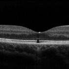
Optical coherence tomography of the left eye of a 60-year-old man with an acquired optic pit maculopathy and glaucoma. The image shows an enlarged optic disc cup and a macular serous detachment.
Photographer: Andree Henaine-Berra. Asociacion Para Evitar la Ceguera en Mexico. Mexico City.
Imaging device: Heidelberg Spectralis
Condition/keywords: glaucoma, maculopathy, optic pit
-
 Acquired Optic Pit Maculopathy
Aug 20 2014 by Andree Henaine-Berra, MD
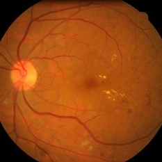
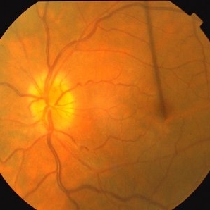
Fundus photograph of the left eye of a 60-year-old man with an acquired optic pit maculopathy and glaucoma. The image shows an enlarged optic disc cup and a macular serous detachment.
Photographer: Andree Henaine-Berra. Asociacion Para Evitar la Ceguera en Mexico. Mexico City.
Imaging device: Heidelberg Spectralis
Condition/keywords: glaucoma, maculopathy, optic pit
-
 Acquired Optic Pit Maculopathy
Aug 20 2014 by Andree Henaine-Berra, MD
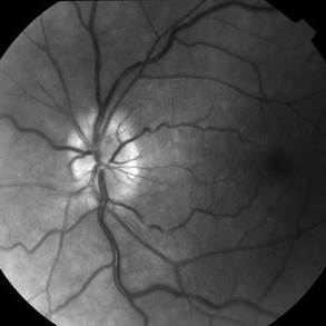
Autofluorescence image of the left eye of a 60-year-old man with an acquired optic pit maculopathy and glaucoma.
Photographer: Andree Henaine-Berra. Asociacion Para Evitar la Ceguera en Mexico. Mexico City.
Imaging device: Heidelberg Spectralis
Condition/keywords: glaucoma, maculopathy, optic pit
-
 Acquired Optic Pit Maculopathy
Aug 20 2014 by Andree Henaine-Berra, MD
Optical coherence tomography of the left eye of a 60-year-old man with an acquired optic pit maculopathy and glaucoma. The image shows subretinal fluid extending to the optic nerve and schisis of the outer retinal layers.
Photographer: Andree Henaine-Berra. Asociacion Para Evitar la Ceguera en Mexico. Mexico City.
Imaging device: Heidelberg Spectralis
Condition/keywords: glaucoma, maculopathy, optic pit
-
 Atrophic Maculopathy
Dec 29 2012 by Barbara Parolini, MD
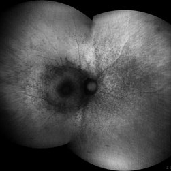
Panoramic autofluorescence picture of a 77-year-old woman with atrophic maculopathy
Photographer: Barbara Parolini, MD
Imaging device: daytona
Condition/keywords: atrophy, maculopathy
-
 Bilateral “Bull's eye”pattern maculopathy
Mar 14 2023 by Anfisa Ayalon, MD
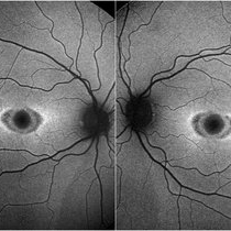
Both eyes fundus autofluorescence image of a 38-year-old female with “Bull's eye” pattern maculopathy. There is no history of medication use associated with retinal toxicity. BCVA RE 20/25+2, LE 20/20-3
Photographer: Danielle Ferguson and Alec Bertoni, University of Pittsburgh Medical Center
Condition/keywords: bull's eye maculopathy, maculopathy, retina
-
 CERKL-related Cone Rod Dystrophy
Jun 27 2022 by Hanna Choi
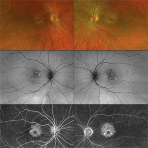
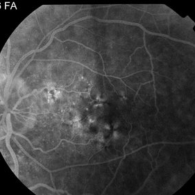
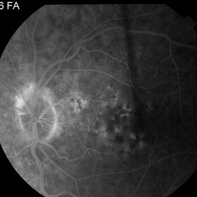
37-year-old female with cone-rod dystrophy. Developed photophobia and progressive blurry vision in the third decade. VA 20/40 OD, 20/30 OS. The patient is compound heterozygous for pathogenic mutations in the CERKL gene (Arg465Trp and Arg283*).
Photographer: Kaitlynn Silva, New England Retina Consultants
Imaging device: Ultrawide-field Optos Fundus Photography, Autofluorescence, Fluorescein Angiography
Condition/keywords: cone dystrophy, inherited retinal disease, maculopathy
-
 Chloroquine maculopathy
Jun 22 2022 by JORGE SOBERANES
Fundus autofluorescence of a bull´s eye maculopathy of a 55-year-old woman treated for ten years with choloquine.
Photographer: Jorge I. Soberanes MD, Asociación para Evitar la Ceguera en México.
Imaging device: Zeiss Clarus 700 (Green autofluorescence)
Condition/keywords: bull's eye maculopathy, chloroquine, fundus autofluorescence (FAF), macula, maculopathy
-
 Elmiron Toxicity
Jan 15 2025 by Virginia Gebhart
54 year old female with pigmentary degeneration secondary to Elmiron. Stippled RPE maculopathy has lightly progressed with stable vision compared to previous visits. BCVA 20/200 OU. Pt reports taking Elmiron from 2010 to 2019.
Photographer: Virginia Gebhart
Imaging device: Optos California
Condition/keywords: autofluorescence imaging, Maculopathy, secondary pigmentary degeneration
-
 Horseshoe Tear in Retinitis Pigmentosa
Mar 22 2021 by ASRS Staff
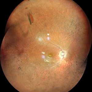
Montage of 25-year-old patient, high myopic patient came with complaint of diminution of vision in both eyes and on posterior segment examination of right eye, HST was present along with maculopathy.
Imaging device: Nidek Mirante
Condition/keywords: maculopathy, retinitis pigmentosa
-
 Laser Pointer Maculopathy
Apr 28 2019 by Bastián Schmidt Arias
Optical coherence tomography of a 13-year-old adolescent with a laser pointer maculopathy, 20/20 visual acuity with paracentral scotoma.
Photographer: Bastian Schmidt
Imaging device: Topcon 3D OCT-2000
Condition/keywords: laser pointer maculopathy, maculopathy, optical coherence tomography (OCT)
-
 Laser Pointer Maculopathy
Jul 1 2018 by John S. King, MD
9-year-old with two month history of decreased vision that has improved some over time. 20/20 OD and 20/30 OS. Further prompting, he admitted to playing with "toy lasers" with his friends recently. He also looked at the eclipse, but used protective eye wear. Photos show some RPE defects characterized by a hyperpigmented center with hypopigmented halo.
Photographer: Karin Aletter
Imaging device: Topcon
Condition/keywords: laser pointer maculopathy, laser pointer retinopathy, maculopathy
-
 Laser Pointer Maculopathy
Jul 1 2018 by John S. King, MD
9-year-old with two month history of decreased vision that has improved some over time. 20/20 OD and 20/30 OS. Further prompting, he admitted to playing with "toy lasers" with his friends recently. He also looked at the eclipse, but used protective eye wear. Photos show small RPE defects characterized by a hyperpigmented center with hypopigmented halo.
Photographer: Karin Aletter
Imaging device: Topcon
Condition/keywords: laser pointer maculopathy, laser pointer retinopathy, maculopathy
-
 Laser Pointer Maculopathy
Jul 1 2018 by John S. King, MD
9-year-old with two month history of decreased vision that has improved some over time. 20/20 OD and 20/30 OS. Further prompting, he admitted to playing with "toy lasers" with his friends recently. He also looked at the eclipse, but used protective eye wear. OCT shows an outer retinal hole.
Photographer: Karin Aletter
Imaging device: Cirrus
Condition/keywords: laser pointer maculopathy, laser pointer retinopathy, maculopathy
-
 Maculopathy and IRMA Left Retina
Dec 18 2013 by Talal basha
Maculopathy and IRMA left retina.
Condition/keywords: intraretinal microvascular abnormalities, maculopathy
-
 Myotonic Dystrophy Maculopathy
Sep 12 2014 by Thomas A. Ciulla, MD, MBA, FASRS
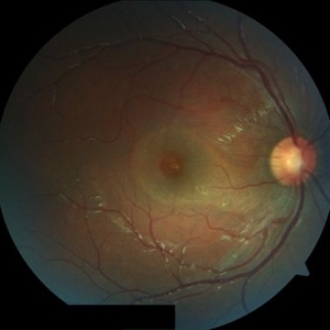
This 43-year-old woman has a history of myotonic dystrophy. Note the subtle pigmentary changes in the macula typical of this disorder. Her visual acuity measured 20/30.
Photographer: Thomas Steele
Condition/keywords: maculopathy
-
 Myotonic Dystrophy Maculopathy
Sep 12 2014 by Thomas A. Ciulla, MD, MBA, FASRS
This 43-year-old woman has a history of myotonic dystrophy. Note the pigmentary changes in the macula typical of this disorder.
Photographer: Thomas Steele
Condition/keywords: maculopathy
-
 Myotonic Dystrophy Maculopathy
Sep 12 2014 by Thomas A. Ciulla, MD, MBA, FASRS
This 43-year-old woman has a history of myotonic dystrophy. Note the pigmentary changes in the macula typical of this disorder. There is no evidence of choroidal neovascularization in this left eye.
Photographer: Thomas Steele
Condition/keywords: maculopathy
-
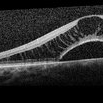
 OCT Myopic Staphyloma With Schisis and ERM
Apr 24 2014 by Scott E. Pautler, MD
OCT of high myope with asymptomatic macular schisis.
Imaging device: Heidelberg Spectralis
Condition/keywords: foveal schisis, maculopathy, maculoschisis, optical coherence tomography (OCT), pathologic myopia, staphyloma
-
---thumb.jpg/image-square;max$300,300.ImageHandler) OCT Optic Pit Maculopathy Post-op
OCT Optic Pit Maculopathy Post-op
Jan 10 2014 by Susanna S. Park, MD, PhD
OCT image taken 1 year after vitrectomy with gas tamponade for macular schisis and detachment and outer lamellar hole associated with optic pit shows normal macular morphology with only mild disruption of the foveal photoreceptor layer.
Photographer: Ellen Redenbo, University of California Davis Eye Center
Condition/keywords: macular schisis, maculopathy, optical coherence tomography (OCT)
-
 Optic Disc Pit Associated with Multilayered Retinoschisis
Apr 26 2023 by Shaleen Arora
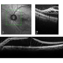
46-year-old female with an optic nerve pit in the left eye (A). OCT reveals retinoschisis involving multiple retinal layers due to intraretinal fluid tracking from the nerve pit (B). Muller cell processes maintain the architecture of individual retinal layers in the region of retinoschisis (C).
Photographer: George Washington University, Department of Ophthalmology
Condition/keywords: maculopathy, optic disc pit, optic pit
-
 Optic Disc Pit Maculopathy
Jan 28 2020 by Pierre-Henry Gabrielle, MD
Optic disc pit maculopathy of a 26-year-old man with optic disc pit maculopathy of his right eye.
Photographer: Pierre-Henry Gabrielle, Ophthalmology department, Dijon University Hospital
Imaging device: Zeiss Visucam
Condition/keywords: fundus photograph, maculopathy, optic disc pit
-
 Optic Disc Pit Maculopathy
Jan 28 2020 by Pierre-Henry Gabrielle, MD
Optic disc pit maculopathy of a 26-year-old man with optic disc pit maculopathy of his right eye.
Photographer: Pierre-Henry Gabrielle, Ophthalmology department, Dijon University Hospital
Imaging device: Heidelberg Spectralis
Condition/keywords: maculopathy, optic disc pit, optical coherence tomography (OCT)
 Loading…