Search results (13 results)
-
 Myopic Traction Maculopathy
Mar 17 2025 by Drew Mitchell
HD 1 line 100x 9 mm scan of a right eye with MTM at stage 3c. Macular Schisis Detachment.
Photographer: Drew Mitchell OCT-C
Imaging device: Zeiss Cirrus 5000
Condition/keywords: full thickness macular hole, Macular hole, myopic foveoschisis, myopic macular schisis, myopic traction maculopathy, PVD
-
 Hypotony Maculopathy
Nov 3 2023 by Matthew Dombrow, MD
31 year old female 4 days s/p Ahmed Valve
Photographer: Cori Sturtevant, Connecticut Retina Consultants, Hamden, Connecticut
Imaging device: Optos - California
Condition/keywords: hypotony maculopathy
-
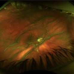
 "The Eye of Sauron"
Mar 14 2023 by Anfisa Ayalon, MD
Fundus autofluorescence image of a 38-year-old female with “Bull's eye” pattern maculopathy. There is no history of medication use associated with retinal toxicity. BCVA RE 20/25+2
Photographer: Danielle Ferguson and Alec Bertoni, University of Pittsburgh Medical Center
Condition/keywords: bull's eye maculopathy, retina
-
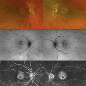 CERKL-related Cone Rod Dystrophy
Jun 27 2022 by Hanna Choi
37-year-old female with cone-rod dystrophy. Developed photophobia and progressive blurry vision in the third decade. VA 20/40 OD, 20/30 OS. The patient is compound heterozygous for pathogenic mutations in the CERKL gene (Arg465Trp and Arg283*).
Photographer: Kaitlynn Silva, New England Retina Consultants
Imaging device: Ultrawide-field Optos Fundus Photography, Autofluorescence, Fluorescein Angiography
Condition/keywords: cone dystrophy, inherited retinal disease, maculopathy
-
 Torpedo Maculopathy
Jul 29 2020 by Yoshihiro Yonekawa, MD, FASRS
Fundus photograph of a 10-year-old boy with an incidentally identified torpedo maculopathy.
Photographer: Suely Bascope
Imaging device: Topcon
Condition/keywords: macula lesion, pediatric retina, torpedo maculopathy
-
 Focal Retinitis- Unilateral Acute Idiopathic Maculopathy OS.
Nov 13 2019 by Tammy Mclaughlin
No prodromal URI. Symptoms present for 1 1/2 weeks. No serous detachment presently. Does have typical circular RPE changes in macula without leakage or edema. Typical course is spontaneous improvement with residual RPE changes. Recommended observation.
Photographer: Tammy Mclaughlin
Imaging device: Zeiss Visucam
Condition/keywords: focal retinitis, unilateral acute idiopathic maculopathy
-
 Ocular Hypotony Due to Leaking Bleb
Apr 1 2019 by Anfisa Ayalon, MD
81-year-old male who had trabeculectomy in his right eye 4 years ago, presented to the emergency room with complains of decreased vision in that eye for two months. Slit-lamp examination showed cystic bleb with leakage, intraocular pressure was 0 MMHg. Fundus examination showed hypotony maculopathy, peripheral choroidal detachments, multiple chorioretinal folds with subretinal fluid.
Photographer: Anfisa Ayalon, MD., Meir Medical Center, Kfar Saba, Israel.
Imaging device: California, Optos 200 DTX
Condition/keywords: choroidal detachment, hypotonous retinopathy, hypotony maculopathy
-
 Laser Pointer Maculopathy
Jul 1 2018 by John S. King, MD
9-year-old with two month history of decreased vision that has improved some over time. 20/20 OD and 20/30 OS. Further prompting, he admitted to playing with "toy lasers" with his friends recently. He also looked at the eclipse, but used protective eye wear. Photos show small RPE defects characterized by a hyperpigmented center with hypopigmented halo.
Photographer: Karin Aletter
Imaging device: Topcon
Condition/keywords: laser pointer maculopathy, laser pointer retinopathy, maculopathy
-
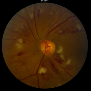 Radiation Retinopathy
Mar 12 2016 by David Callanan, MD
55-year-old with background diabetic retinopathy that developed renal cell carcinoma. Underwent radiation to left orbit.
Condition/keywords: radiation maculopathy
-
 Chorioretinitis Sclopetaria
Jan 22 2016 by Jorge Morales-Martínez, MD
Fundus photograph of a 27-year-old male that sustained a traumatic injury in his left eye with a paintball projectile. Fundus examination showed a large subretinal hemorrhage, areas of commotio retinae and maculopathy.
Photographer: Jorge Morales-Martínez MD
-
 OCT Myopic Staphyloma With Schisis and ERM
Apr 24 2014 by Scott E. Pautler, MD
OCT of high myope with asymptomatic macular schisis.
Imaging device: Heidelberg Spectralis
Condition/keywords: foveal schisis, maculopathy, maculoschisis, optical coherence tomography (OCT), pathologic myopia, staphyloma
-
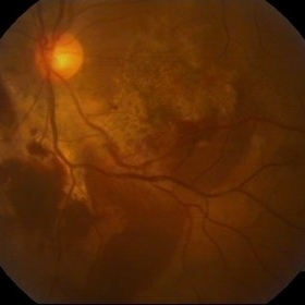
---thumb.JPG/image-square;max$300,300.ImageHandler) Optic Pit Maculopathy - Fundus Photograph
Optic Pit Maculopathy - Fundus Photograph
Oct 14 2013 by Cagri G Besirli, MD, PhD, FASRS
10-year-old girl with congenital optic pit and recent vision loss secondary to optic pit maculopathy.
Imaging device: Optos
Condition/keywords: fundus photograph, maculopathy
-
 Polymorphous Vitelliform Maculopathy
Jul 11 2013 by Eric M. Shrier, DO
38-year-old Middle Eastern female referred for IFN screening (Hep. c +).
Condition/keywords: idiopathic, polymorphous exudative vitelliform maculopathy
 Loading…