Search results (561 results)
-
 BCAMD FP
May 13 2025 by Moazzam Parvez
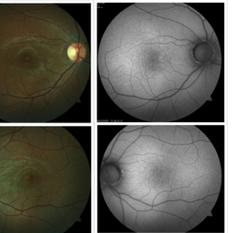
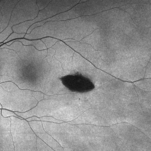
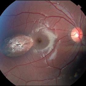
Fundus photograph of a 40 year old woman with a bulls eye maculopathy
Photographer: Dr Moazzam Parvez, Netralayam , Kolkata
Imaging device: Topcon Maestro 2
Condition/keywords: Benign concentric annular macular dystrophy
-
 BCAMD AF
May 13 2025 by Moazzam Parvez
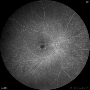
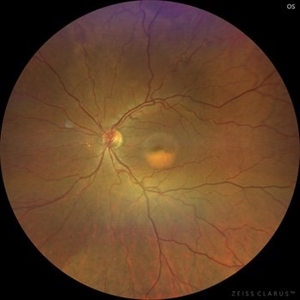
Auto fluorescence image of the right eye with a bulls eye maculopathy depicting central auto hypo fluorescence surrounding a relatively normal fovea.
Photographer: Dr Moazzam Parvez , Netralayam, Kolkata
Imaging device: Heidelberg Spectralis
Condition/keywords: bulls eye maculopathy
-
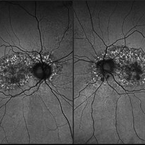
 Bilateral Benign Yellow Dot Maculopathy
May 6 2025 by Amol yuvraj ganvir
A 37-year-old female patient presented for a routine eye examination. Her best-corrected visual acuity was 6/6 in both eyes. Fundus examination revealed multiple small yellow dots over the macula in both eyes. FAF imaging demonstrated characteristic hyperautofluorescence corresponding to these dots.
Photographer: Dr. Amol Ganvir, Vitreo-Retina Fellow, Ishwar Eye Centre, Rohtak, Haryana
Imaging device: Visucam-Zeiss
Condition/keywords: Autoflourescence, yellow dots
-
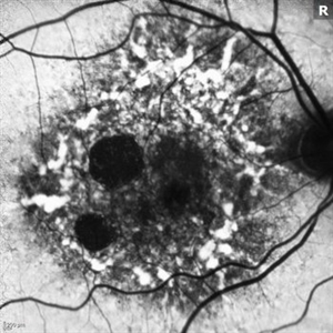
 Toxic Maculopathy (Elmiron)
Apr 9 2025 by Virginia Gebhart
79 year old male with toxic maculopathy from long term use of Elmiron (15+ yrs.) On exam there is stippled RPE changes, pigment clumping, and subretinal deposits. BCVA 20/100 | 20/40.
Photographer: Virginia Gebhart, Retina Consultants of Carolina
Imaging device: Optos California
Condition/keywords: autofluorescence imaging, cystoid macular degeneration, Elmiron Toxicity, Toxic Maculopathy
-
 Elmiron Toxicity
Mar 25 2025 by Toolie Winters
Fundus autofluorescence image of a 69-year-old woman with toxic maculopathy OU due to Elmiron usage. Patient stopped using Elmiron in the late 2010s after having been on it for 17 years. The patient has areas of outer retinal and RPE atrophy temporal to fovea that have expanded compared to photos from two years ago. At the time of this appointment, her VA OD was sc20/40-1+2 PH20/30 and VA OS was scCF @ 1 foot.
Photographer: Toolie Winters
Imaging device: Heidelberg Spectralis
Condition/keywords: Elmiron Toxicity, FAF, fundus autofluorescence (FAF), Heidelburg Spectralis, Pentosan Toxicity, Toxic Maculopathy
-
 Myopic Traction Maculopathy
Mar 17 2025 by Drew Mitchell
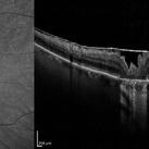
HD 1 line 100x 9 mm scan of a right eye with MTM at stage 3c. Macular Schisis Detachment.
Photographer: Drew Mitchell OCT-C
Imaging device: Zeiss Cirrus 5000
Condition/keywords: full thickness macular hole, Macular hole, myopic foveoschisis, myopic macular schisis, myopic traction maculopathy, PVD
-
 Elmiron Toxicity
Jan 15 2025 by Virginia Gebhart
54 year old female with pigmentary degeneration secondary to Elmiron. Stippled RPE maculopathy has lightly progressed with stable vision compared to previous visits. BCVA 20/200 OU. Pt reports taking Elmiron from 2010 to 2019.
Photographer: Virginia Gebhart
Imaging device: Optos California
Condition/keywords: autofluorescence imaging, Maculopathy, secondary pigmentary degeneration
-
 Pigmentary Degeneration of Retina (Secondary to Elmiron)
Nov 27 2024 by Virginia Gebhart
77 year old female with advanced geographic atrophy after years of Elmiron use (stopped in 2018). Serial exams show continued progression of GA. Central vision limited, vision remains stable and patient does not report noticing any changes.
Photographer: Virginia Gebhart, Retina Consultants of Carolina
Imaging device: Optos California
Condition/keywords: geographic atrophy, secondary pigmentary degeneration, toxic maculopathy
-
 Proliferative Diabetic Retinopathy with Neovascularization
Nov 15 2024 by Júlio Andrade
Fundus photograph of a patient with proliferative diabetic retinopathy presenting with extensive neovascularization, microaneurysms, and evidence of fluid leakage. Key findings include the presence of new blood vessel formation, indicative of advanced retinal ischemia and disease progression.
Photographer: úlio Andrade, Retina Instituto, Belo Horizonte
Imaging device: Zeiss Clarus 700
Condition/keywords: diabetic blindness, ischaemic diabetic maculopathy, Neovascularisation elsewhere (NVE), NEOVASCULARISATION OF DISC, Proliferative Diabetic retinopathy
-
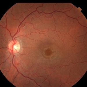
 Best Disease
Nov 7 2024 by Virginia Gebhart
Fluorescein angiogram of 49 year female with Best Disease. Genetic testing done in 2000 confirms Best Disease and also possible Stargardts mutation. Characteristic bullseye maculopathy with surrounding yellowish flecks are present in both eyes.
Photographer: Virginia Gebhart, Retina Consultants of Carolina
Imaging device: Optos California
Condition/keywords: Best disease, fluorescein angiogram (FA)
-
 Torpedo Retinopathy
Oct 31 2024 by AVIK DEY SARKAR, MS, FVRS, FAICO(VR)
This is a 42 year old male with known history of diabetes mellitus for past 10 years. Patient presented with complains regarding presbyopia. On dilated fundoscopy, along with dot and blot hemorrhages, in the infero-temporal near-periphery outside the vascular arcade a hypopigmented torpedo-shaped lesion was noted. On OCT, outer retinal attenuation with sublesional choriocappilaris layer thinning was noted. The lesion is diagnosed as torpedo retinopathy. Torpedo maculopathy is rare in clinical practice and usually is found at the margin of temporal arcade over "Temporal Bulge". But this lesion is seen well away from the posterior pole. This case indicates the necessity of substituting the terminology "Torpedo Maculopathy" with "Torpedo Retinopathy" as mentioned earlier in ophthalmic literature.
Photographer: Dr. Avik Dey Sarkar, MBBS, MS, FVRS, FAICO, Consultant, Department of Vitreoretinal Services, Aravind Eye Hospital, Madurai, India
Imaging device: Wide angled Fundus imaging with Clarus 300
Condition/keywords: torpedo maculopathy, torpedo Retinopathy
-
 Hidroxychloroquine Retinopathy
Aug 27 2024 by Monica Elena Cortizo Brown , MD
72 year old female with long-term use of hydroxichloroquine for more than 5 years with parafoveal retinal pigment atrophy and “Bull's eye maculopathy”
Photographer: Mónica Elena Cortizo, Hospital de la Luz, Ciudad de México
Condition/keywords: Hidroxychloroquine maculopathy, Medication toxicity
-
 Morning glory disc anomaly-associated maculopathy: fibroglial tissue with a Mac-Off serous retinal detachment.
Jun 26 2024 by JULIAN VILLARREAL, MD
19 year old with a Morning glory disc anomaly-associated maculopathy: fibroglial tissue with a Mac-Off serous retinal detachment.
Photographer: Julián Villarreal MD
Imaging device: Mirante
Condition/keywords: fibroglial tissue, Morning Glory Anomaly, retinal detachment of the macula
-
 ILM Peeling in Case of Optic Disc Pit Maculopathy
Jun 14 2024 by Tejaswita Verma
Intraoperative still of a 38 year old male post initiation of ILM peeling in a case of optic disc pit maculopathy.
Photographer: DR. TEJASWITA VERMA
Condition/keywords: intraoperative, optic pit
-
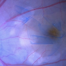
 PAMM
PAMM
May 24 2024 by Gustavo Del Castillo-Marquez, MD
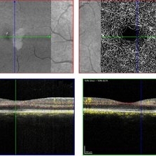
EN FACE OCT ANGIOPLEX ELITE image of an 44-year-old man with Paracentral Acute Middle Maculopathy of early onset.
Photographer: Gustavo Del Castillo-Márquez, Asociación Para Evitar la Ceguera en México, CDMX
Imaging device: Zeis Ciruss Angioplex 5000
Condition/keywords: enface imaging, PAMM
-
 Torpedo Maculopathy
May 17 2024 by Júlio Andrade
Patient, 12 years old, routine, 20/20 vision in both eyes. A surprise in the fundoscopy of the left eye: a hypopigmented area in the shape of a torpedo inferior temporal to the macula, pointing towards the fovea with a hyperpigmented tail, and another similar lesion inferior to the first without a hypopigmented area. An OCT was performed, showing choroidal cavitation. We continue to follow up, after 3 years the condition remains stable, without the development of neovascularization.
Photographer: Tereza Kanadani
Condition/keywords: maculopathy, OCT, Torpedo Maculopathy
-
 Torpedo Maculopathy
Feb 20 2024 by Soobien Lee
Optos color fundus photograph of a 35-year-old asymptomatic female with no ocular or medical history with stable and chronic appearing torpedo-shaped macula lesion in the left eye.
Photographer: Peter Sotirakos, Elman Retina Group
Imaging device: Optos Ultra-Widefield Imaging
Condition/keywords: macula, Optos, torpedo maculopathy
-
 Torpedo Maculopathy
Feb 20 2024 by Soobien Lee
Optos fundus autofluorescence photograph of a 35-year-old asymptomatic female with no ocular or medical history with stable and chronic appearing torpedo-shaped macula lesion in the left eye.
Photographer: Peter Sotirakos, Elman Retina Group
Imaging device: Optos Ultra-Widefield Autoflurescence Imaging
Condition/keywords: autofluorescence imaging, genetics, macula, maculopathy, Optos, torpedo maculopathy
-
 Flourescein Angiography of Cloroquine Toxicity
Feb 12 2024 by BENITO VERGARA, MD
Image of a late phase fluorescein angiography at 4 minutes and 30 seconds of a 58-year-old woman treated with chloroquine at a daily dose of 3mg/kg, (recommended dose >2.3 mg/kg) that shows circular window defect suggestive of bullseye maculopathy.
Photographer: Benito Vergara Flores.
Imaging device: Clarus 700
Condition/keywords: chloroquine toxicity
-
 Dislocated Iol With Hypotony Maculopathy and Hemorrhagic Choroidal
Feb 9 2024 by Sandra R Montezuma, MD
28 year old year-old male with history of congenital cataract of the right eye, s/p cataract extraction in 1999, s/p lens implant in 2011, presented with a dislocated IOL, hypotony, retina folds, hypotony maculopathy and hemorrhagic nasal choroidal after unsuccessful surgery to attempt remove the dislocated lens.
Photographer: Scott Baker, University of Minnesota
Condition/keywords: choroidals, dislocated posterior chamber intraocular lens (PCIOL), hypotony maculopathy, retina folds
-
 Bullseye Maculopathy
Jan 22 2024 by Kali Jend
Optical coherence tomography of a 73-year-old female with Bullseye Macular Changes affecting her left eye. Patient reports having a family history of this condition and denies prior Plaquenil or Elmiron use. Compared to previous imaging, the patient's condition progressed in the left eye from 2020 to 2023. Patient has a history of fluctuating Diabetic Macular Edema and a current Epiretinal Membrane as well. Patient's vision was Ncc20/60 at the time the image was taken.
Photographer: Kali Jend
Imaging device: Heidelberg Spectralis
Condition/keywords: bullseye maculopathy, epiretinal membrane (ERM), heidelberg spectralis, left eye, macular pucker, OCT, optical coherence tomography (OCT)
-
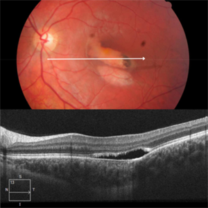
Paracentral Acute Middle Maculopathy
Nov 29 2023 by Daniel Davis, OCT-C
30 yo female OCT with Paracentral Acute Middle Maculopathy (PAMM) OD VA OD: sc20/60+1
Condition/keywords: OCT, PAMM
-
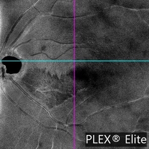
 PAMM-OCTA
Nov 29 2023 by Daniel Davis, OCT-C
OCT-A of a 30 yo female with PAMM OD.
Photographer: Daniel Davis, OCT-C
Imaging device: Heidelberg Spectralis
Condition/keywords: OCTA, paracentral acute middle maculopathy
-
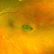
 Torpedo Maculopathy
Nov 25 2023 by Neeket R. Patel, MD
Fundus photograph of a 12-year-old female with an incidental finding of torpedo maculopathy.
Condition/keywords: torpedo maculopathy
-
 Adult-onset foveomacular vitelliform dystrophy
Nov 10 2023 by Josue Elias Guevara Cruz
Fundus photograph of an 54 year-old woman with past history of unknown maculopathy.
Photographer: Josue Guevara. UNAM
Imaging device: Zeiss Clarus
Condition/keywords: adult foveomacular dystrophy
 Loading…