Search results (176 results)
-
 Proliferative Diabetic Retinopathy with Choroidal Effusion Status Post PRP
Dec 15 2020 by Manish Nagpal, MD, FRCS (UK), FASRS
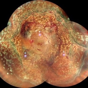
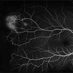
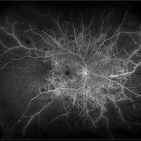
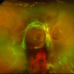
A 17-year-old juvenile diabetic patient came to us with extensive neovascular proliferations and PRP done a week back and had developed 360 degree choroidal effusion as seen in this wide field montage image
Photographer: Sham Talati, Retina Fellow , Retina Foundation, Ahmedabad, India
Imaging device: Mirante CSLO
Condition/keywords: choroidal effusion, diabetic retinopathy, proliferative diabetic retinopathy (PDR)
-
 PRP laser
Mar 29 2013 by Henry J. Kaplan, MD
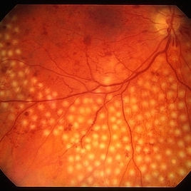
Right after PRP laser in PDR.
Condition/keywords: laser photocoagulation, pan-retinal photocoagulation (PRP)
-
 Rod Cone dystrophy
Nov 29 2022 by Niloofar Piri, MD
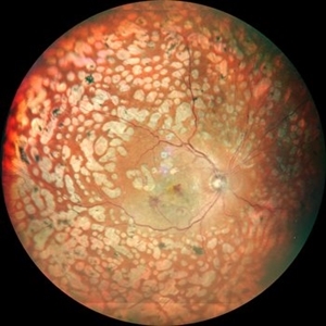
Fundus photograph of the left eye in a 58 yo male with rod cone dystrophy. He presented with night blindness and peripheral vision loss since youth and recent decrease in central vision for the past 10 years. Notice waxy pallor of the nerve, severe arterial narrowing and chorioretinal atrophy mainly around the arcades as well as posterior pole along with RPE hyperplastic changes and atrophy. RPE atrophy in midperiphery has coin shaped appearance. FAF has characteristic appearance (uploaded separately) He has one pathogenic variants of both CEP290 and PRPH2 genes.
Photographer: Sean Kelso, Saint Louis University
Condition/keywords: hereditary retinal deg, hereditary retinal dystrophy, Rod cone dystrophy
-
 Rod Cone dystrophy
Nov 29 2022 by Niloofar Piri, MD
Fundus autofluorescence of the left eye in a 58 yo male with rod cone dystrophy. He presented with night blindness and peripheral vision loss since youth and recent decrease in central vision for the past 10 years. Notice multiple coin shaped hypoautofluorescent pacthes within central 20 degrees which are coalescing centrally. (fundus photo uploaded separately) He has one pathogenic variants of both CEP290 and PRPH2 genes.
Photographer: Sean Kelso, Saint Louis University
Condition/keywords: hereditary retinal degeneration, hereditary retinal dystrophy, rod cone dystrophy
-
 Proliferative Sickle Cell Retinopathy
Jan 29 2021 by Olivia Rainey
Ultra-widefield fluorescein angiogram of a 24-year-old female with proliferative sickle cell retinopathy affecting her right eye. The physician's interpretation of the fluorescein shows seafan neovascularization superotemporally, AV anastomeses, and good peripheral laser. He performed scatter PRP OD on 12/2/2020 to nonperfusion in temporal far periphery. The patient's 12/2020 Hb electrophoresis came back showing Hb SC (rather than sickle cell trait). Patient was born at full term, but she reports that her mother used drugs while pregnant with the patient. The patient also mentioned that her niece has full sickle cell disease and her grandmother, mother, and sibling all have condition on the sickle cell spectrum.
Photographer: Olivia Rainey, OCT-C, COA
Imaging device: Optos California
Condition/keywords: fluorescein angiogram (FA), fluorescein leakage, neovascularization (NV), neovascularization elsewhere (NVE), Optos, sea fan, sickle cell retinopathy
-
 Retinitis Pigmentosa with PPRPE
Jan 27 2025 by Vishal Agrawal, MD, FRCS,FACS,FASRS
16 year-old male patient presented with DOV, nyctalopia and nystagmus. Fundus revealed pigment clumping, pale disc and preserved para-arteriolar retinal pigment epithelium (PPRPE) in both eyes. Genetic testing revealed CRB1 gene mutation.
Photographer: Dr Ayushi
Imaging device: Clarus 700
Condition/keywords: retinitis pigmentosa
-
 Retinopathy of Prematurity
Aug 26 2021 by Stefanie Palmer
Patient has neovascular ridge temporal with elevation of vessels above the PRP scars. The image was obtained with the flying baby technique.
Photographer: Stefanie Palmer, CRA
Condition/keywords: retinopathy of prematurity (ROP)
-
 Retinopathy of Prematurity
Aug 26 2021 by Stefanie Palmer
Patient has neovascular ridge temporal with elevation of vessels above the PRP scars. The image was obtained with the flying baby technique.
Photographer: Stefanie Palmer, CRA
Condition/keywords: retinopathy of prematurity (ROP)
-
 Sickle Cell Retinopathy
Feb 15 2021 by Kim Barrett
24-year-old female with Sickle Cell Retinopathy, stage 3. She confirms she has the trait as well as her grandmother, mother and a sibling. She has seafan neovascularization superotemporal OD. Current VA is 20/20. Photo is pre-PRP laser with areas of non-profusion temporally.
Photographer: Kim Barrett C.O.A. Retina Specialist of Michigan, Grand Rapids, MI
Imaging device: Optos California
Condition/keywords: neovascularization (NV), pan-retinal photocoagulation (PRP), sickle cell retinopathy, stage 3, trait
-
 Active diabetic retinopathy despite PRP
Oct 30 2022 by Diego Andrés Rodriguez, MD
A 52-year-old patient with active proliferative diabetic retinopathy despite good glycemic control and PRP performed 1 year ago in the right eye
Photographer: Sociedad de Cirugía Ocular
Imaging device: Clarus 700
Condition/keywords: diabetic retinopathy, pan-retinal photocoagulation (PRP), proliferative diabetic retinopathy (PDR), wide angle imaging
-
 Branch Retinal Vein Occlusion
Dec 9 2020 by Olivia Rainey
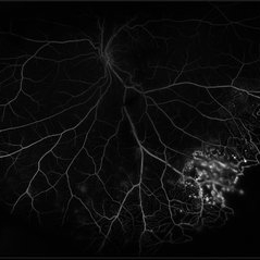
Ultra-widefield angiogram of a 78-year-old male with a branch retinal vein occlusion affecting his right eye. The patient was diagnosed on 12/17/12 at another practice. The physician noted that there wasn't NVE noted, however areas of micoaneurysmal dilation is present. She noted retinal ischemia secondary to BRVO. 12/8/20 leakage on FA noted to be worsening compared to his previous angiography. She has concern for progressing NVE and recommends sector PRP after injection of antiVEGF series of 3 for the health of the eye.
Photographer: Olivia Rainey, OCT-C, COA
Imaging device: Optos California
Condition/keywords: branch retinal vein occlusion (BRVO), macular branch retinal vein occlusion (BRVO), non-perfusion, scleral buckle, vitreoretinal surgery
-
 Central Retinal Artery Occlusion
Apr 20 2018 by Kim Barrett
64-year-old female woke with no vision in her right eye. This image was taken at 6:11 minutes and the vessels have not filled. Patient has been treated with PRP laser and anti-VEGF therapy. Current vision is CF @ 2 ft.
Photographer: Kim Barrett C.O.A.
Imaging device: Heidelberg
Condition/keywords: central retinal artery occlusion (CRAO), diabetes, hypertension, smoker, uncontrolled
-
 Central Retinal Vein Occlusion with Retinal Neovascularization
Jan 19 2022 by Olivia Rainey
Ultra-widefield fluorescein angiogram of a 56-year-old male with a Central Retinal Vein Occlusion with Retinal Neovascularization affecting his left eye. The patient presented on 1/19/2022 with scNLP vision in the left eye. The patient has good PRP, however areas of ischemia still remain untreated by laser. He also has severe neovascular glaucoma contributing to his poor vision.
Photographer: Olivia Rainey, OCT-C, COA
Imaging device: Optos California
Condition/keywords: central retinal vein occlusion (CRVO), FA early phase, fluorescein angiogram (FA), hemorrhage, ischemic CRVO, left eye, neovascular glaucoma, Optos, pan-retinal photocoagulation (PRP), retinal ischemia, retinal neovascularization, ultra-wide field imaging
-
 Central Retinal Vein Occlusion with Severe Retinal Ischemia
Jan 19 2022 by Olivia Rainey
Ultra-widefield fluorescein angiogram of a 56-year-old male with a Central Retinal Vein Occlusion with Severe Retinal Ischemia affecting his right eye. The patient presented on 1/19/2022, sc20/20-2 vision in the right eye. The patient has had a good response to Eylea with complete resolution of edema. The physician is considering PRP to ischemic periphery in the future and given the degree of ischemia in both eyes, she recommends that the patient's PCP check carotid Doppler US.
Photographer: Olivia Rainey, OCT-C, COA
Imaging device: Optos California
Condition/keywords: central retinal vein occlusion (CRVO), FA late phase, fluorescein angiogram (FA), ischemic CRVO, Optos, retinal ischemia, ultra-wide field imaging
-
 CNVM in Pan-retinal Photocoagulated Patient
Dec 30 2020 by ASRS Staff
Wide fundus photograph of 65-year-old, female, diabetic patient.
Imaging device: Nidek Mirante
Condition/keywords: age-related macular degeneration (AMD), diabetes, pan-retinal photocoagulation (PRP)
-
 Coats' Disease
Jul 16 2019 by Kim Barrett
Ultra-wide field fluorescein angiogram of a 23-year-old male with Coats' disease, presented with distorted vision affecting his left eye. He reported seeing flashes and floaters since January of 2019, but the flashes had resolved. He was treated with Intravitreal Preservative Free Triamcinolone in the office and scheduled for PRP laser in the near future.
Photographer: Kim Barrett
Imaging device: Optos
Condition/keywords: Coats' disease, fluorescein angiogram (FA), fluorescein leakage, inferior retina, ischemia, left eye, Optos, ultra-wide field imaging
-
 Coats' Disease
Jul 10 2018 by Karen Panzegrau
Ultra-wide field images of a 30-year-old male with Coats' Disease affecting his right eye. Patient had sectoral PRP with significant improvement in lipid after 8 months of being lost to folllow up. Vision has improved beyond expectations given severity of lipid.
Photographer: Karen Panzegrau
Imaging device: Optos
Condition/keywords: Coats' disease, fundus photograph, lipid exudation, pan-retinal photocoagulation (PRP)
-
 Diabetic Retinopathy
Oct 18 2012 by Raj K. Maturi, MD
Photographer: Tom Steele, CRA
Imaging device: Optos
Condition/keywords: pan-retinal photocoagulation (PRP)
-
 Diabetic Retinopathy
Oct 18 2012 by Raj K. Maturi, MD
Photographer: Tom Steele, CRA
Imaging device: Optos
Condition/keywords: pan-retinal photocoagulation (PRP)
-
 Diabetic Retinopathy Treated with PRP Laser
Jun 8 2021 by Ronald Coriasso
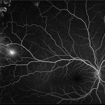
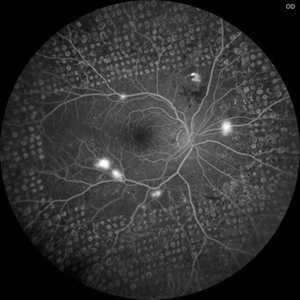
Diabetic retina treated with complete 360 PRP laser, taken during fluorescein angiogram.
Photographer: Ronald Coriasso
Imaging device: OPTOS
Condition/keywords: pan-retinal photocoagulation (PRP)
-
 Diabetic Tractional Retinal Detachment
Jan 23 2019 by Olivia Rainey
Ultra-wide field pseudocolor image of an 43-year-old female with a diabetic tractional retinal detachment and a vitreous hemorrhage affecting her right eye.
Photographer: Olivia Rainey
Imaging device: Optos
Condition/keywords: diabetes, diabetic traction detachment, Optos, pan-retinal photocoagulation (PRP), proliferative diabetic retinopathy (PDR), pseudocolor, ultra-wide field imaging, vitreous hemorrhage
-
 Extensive Pan-Retinal Photocoagulation
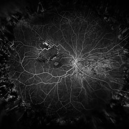
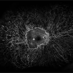
Apr 19 2013 by Brandon G. Busbee, MD
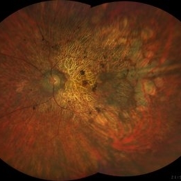
Extensive pan-retinal photocoagulation.
Photographer: Alecia Camp, CRA - Tennessee Retina - Nashville, TN
Imaging device: Topcon TRC 50-EX
Condition/keywords: neovascularization (NV), pan-retinal photocoagulation (PRP)
-
 Exudative Macular Detachment After Intensive Laser Photocoagulation
Mar 12 2016 by Sjakon G Tahija, MD
Fundus photograph of 44-year-old man with exudative detachment of the macula after vitrectomy and ILM peeling for proliferative diabetic retinopathy combined with intensive endolaser photocagulation.
Photographer: Avris Siahaan, Klinik Mata Nusantara
Condition/keywords: exudative detachment, pan-retinal photocoagulation (PRP)
-
 Moyamoya: FA 2 Min OD of an Acute CRAO with CRA Sparing
Nov 17 2019 by John S. King, MD
60-year-old white female presented with five days of acute vision loss in the right eye. She was seen initially by referring doctor after hours five days ago and diagnosed with a CRAO and sent to ED to be evaluated stroke team. Right ICA was 100% closed but completely bypassed. She called four days later c/o redness and eye pain; at this point prominent iris vessels were seen, and she was sent to us. Her background history includes a diagnosis of moyamoya (underwent bilateral cerebral artery bypass 2015); atorvastatin for hypercholesterolemia; ASA; no hx of HTN or heart disease. She had a scleral buckle repair OD in 2017 and later developed a thick ERM, which was repaired in 2018; on her previous visit her acuity was noted at 20/40. On presentation her visual acuity was HM OD and 20/15 OS. IOP was 8 OD and 10 OS. There were prominent iris vessels in the right eye, no cell or flare, and an IOL. The posterior segment exam showed diffuse retinal whitening with attenuated vessels and boxcarring; there was sparing retinal whitening in a central area of the macula that appeared to be supplied by a cilio-retina artery. The FA showed very slow filling of the retinal vessels; there was some early perfusion secondary to the cilio-retinal artery. At 7 minutes there was still significant areas of non-perfusion, as well as macular ischemia. Avastin was administered, and one week later, PRP was performed. On the day PRP was performed, the irregular iris vessels had regressed completely. She said that she had a "sliver" of vision centrally in that eye; her acuity was CF 2' and IOP 12.
Photographer: Gretchen Harper
Imaging device: Topcon
Condition/keywords: central retinal artery occlusion (CRAO), cilioretinal sparing, moyamoya, neovascularization of iris (NVI)
-
 Multifocal Pattern Dystrophy
Feb 5 2025 by Kimberly Wakester
Optomap RGB and AF photograph of an 37-year-old woman with multifocal pattern dystrophy in both eyes. Previously believed to be Stargardts, but genetic testing returned positive for PRPH2 mutation. Likely Multifocal Pattern Dystrophy given phenotypical appearance of SGD. There is stable NVE in the left eye. Will continue to monitor both eyes and consider treatment with PRP laser if needed for NVE in the left eye.
Photographer: Kimberly Wakester, COA
Imaging device: Optos California
Condition/keywords: multifocal pattern dystrophy, NVE, PRPH2 Positive
 Loading…