Search results (176 results)
-
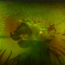
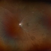
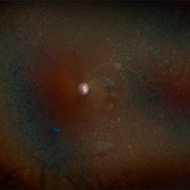
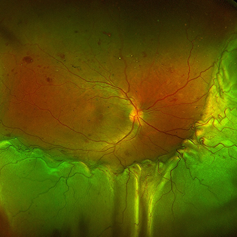
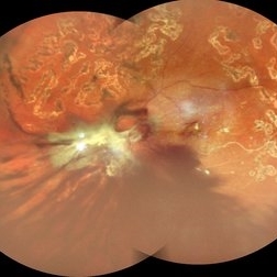 Table Top Tractional Retinal Detachment With Vitreous Hemorrhage in a Case of Proliferative Diabetic Retinopathy
Sep 12 2025 by Akansha Sharma
Color fundus photograph of a 56 year old male with table top tractional retinal detachment with vitreous hemorrhage in a case of proliferative diabetic retinopathy.
Photographer: DR. AKANSHA SHARMA
Condition/keywords: pan-retinal photocoagulation (PRP), PDR, proliferative diabetic retinopathy (PDR), PRP, TABLE TOP TRD, tractional retinal detachment, TRD, VH, vitreous hemorrhage
-
 Vasoproliferative Tumor (FEVR) s/p PPV/PRP
Aug 27 2025 by Virginia Gebhart
39 year old female with an amelanotic vascular lesion inferotemporal with CR atrophy inferior edge and likely lipid exudate superior edge. Pt presented with vitreous and sub-hyaloid hemorrhage. Findings from exam, ultrasound, FA all consistent with FEVR, stage 2. PPV with PRP performed, pt vison has improved from CF@2ft at initial visit to 20/100 PH 20/60 at 1 week post-op. Pt's 2 children have been recently examined with identical findings of FEVR
Photographer: Virginia Gebhart, Retina Consultants of Carolina
Imaging device: Optos California
Condition/keywords: familial exudative vitreoretinopathy (FEVR), pan-retinal photocoagulation (PRP), Vasoproliferative Tumor
-
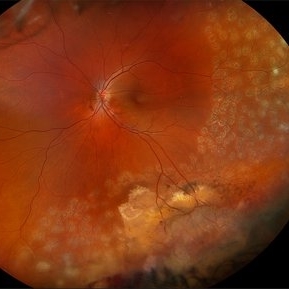
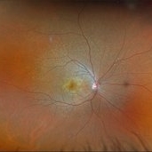
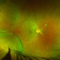
 Proliferative Diabetic Retinopathy
Aug 11 2025 by Marin Shehata
Fundus photograph of a 63 year-old male with diabetic retinopathy has been treated with PRP.
Photographer: Marin Shehata, Retina Consultants of Carolina
Imaging device: Optos California
Condition/keywords: proliferative diabetic retinopathy (PDR), PRP
-
 Retinal Aneurysms
Aug 6 2025 by Korey Starkey
54 year-old patient presents with scattered peripheral aneurysms with exudates. FA was performed showing peripheral nonperfusion and aneurysms. Treated patient with PRP and focal laser to aneurysms and continued observation.
Photographer: Kore Starkey
Imaging device: Optos
Condition/keywords: aneurysm, branch retinal vein occlusion (BRVO), chorioretinal scar, circinate ring, exudates, fundus photography, lesion, Optos, retinal aneurysms
-
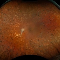
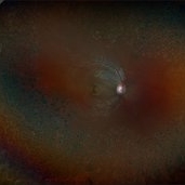
 Unstable PDR s/p Laser
Aug 4 2025 by Anjana Mirajkar, MS Ophthalmology
Fundus photograph of a 60 year old male with an unstable PDR showing traction at the posterior pole with sub hyaloid hemorrhage. Peripheral PRP marks can be seen.
Photographer: Dr. Anjana Mirajkar- HV Desai eye hospital ,Pune
Imaging device: Optos
Condition/keywords: pan-retinal photocoagulation (PRP), proliferative diabetic retinopathy (PDR), subhyaloid hemorrhage, tractional retinal detachment
-
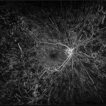
 Ocular Ischemic Syndrome
Jun 18 2025 by Korey Starkey
58-year-old patient with OIS in both eyes. Patient has had PRP in the past, however, presence of NVD with peripheral nonperfusion remains despite PRP.
Photographer: Korey Starkey
Imaging device: Optos
Condition/keywords: DME, FA early phase, fluorescein angiogram (FA), NVD, ocular ischemic syndrome, ois, Optos, peripheral retinal nonperfusion
-
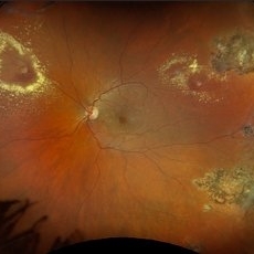
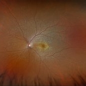
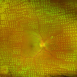
 Proliferative Diabetic Retinopathy S/P Pan Retinal Photocoagulation
Mar 4 2025 by Prithvi Chandrakanth
A 52-year-old female patient presented with complaints of diminishing vision, compounded by uncontrolled diabetes mellitus. Her Fundus examination revealed proliferative diabetic retinopathy, characterized by neovascularization of the disc and elsewhere, and sclerosed vessels. To address this, Pan Retinal Photocoagulation was performed, and the condition stabilized, halting the progression of the disease.
Photographer: DR PRITHVI CHANDRAKANTH, DR CHANDRAKANTH NETHRALAYA, KOZHIKODE, KERALA, INDIA
Imaging device: EIDON
Condition/keywords: Diabetic Retinopathy, Neovascularisation at the Disc (NVD), neovascularization of the disc (NVD), NVD, pan-retinal photocoagulation (PRP), PDR, PDR with NVE (periphery), PRP
-
 Sickle Cell Retinopathy
Feb 24 2025 by Kimberly Wakester
Optomap RGB image of an 24-year-old woman with sickle cell retinopathy in both eyes. There is overall progression of the ischemic vessels and vascular drops out compared to previous images completed in 2021. Oral FA was completed and shows possible progression of peripheral non-perfusion but difficult to determine due to drinking FA dye and images not being as bright. On Clinical exam there is no evidence of NV, RD, or RT in either eye. Patient understands the need for continued follow up care and the likely need for PRP laser in both eyes.
Photographer: Kimberly Wakester, COA
Imaging device: Optos California
Condition/keywords: sickle cell retinopathy
-
 Multifocal Pattern Dystrophy
Feb 5 2025 by Kimberly Wakester
Optomap RGB and AF photograph of an 37-year-old woman with multifocal pattern dystrophy in both eyes. Previously believed to be Stargardts, but genetic testing returned positive for PRPH2 mutation. Likely Multifocal Pattern Dystrophy given phenotypical appearance of SGD. There is stable NVE in the left eye. Will continue to monitor both eyes and consider treatment with PRP laser if needed for NVE in the left eye.
Photographer: Kimberly Wakester, COA
Imaging device: Optos California
Condition/keywords: multifocal pattern dystrophy, NVE, PRPH2 Positive
-
 Systemic Lupus Erythematosus (SLE) Vasculitis
Jan 29 2025 by Kimberly Wakester
Fundus photographs of an 13-year-old boy with Systemic Lupus Erythematosus (SLE) Vasculitis in both eyes s/p PRP laser. Patient is doing well s/p PRP Laser OU and with continued use of oral medications. Patient will be monitored with follow up exams to check for recurring vasculitis or recurring/worsening NVE/NVD. Patient is to continue ongoing management with Rheumatologist.
Photographer: Kimberly Wakester, COA
Imaging device: Optos California
Condition/keywords: NVD, NVE, occlusive vasculitis, pan-retinal photocoagulation (PRP), Systemic Lupus Erythematosus (SLE) Vasculitis
-
 Retinitis Pigmentosa with PPRPE - FAF-G
Jan 27 2025 by Vishal Agrawal, MD, FRCS,FACS,FASRS
16 year-old male patient presented with DOV, nyctalopia and nystagmus. Fundus revealed pigment clumping, pale disc and preserved para-arteriolar retinal pigment epithelium (PPRPE) in both eyes. Genetic testing revealed CRB1 gene mutation.
Photographer: Dr Ayushi Gupta
Imaging device: Clarus 700
Condition/keywords: retinitis pigmentosa
-
 Retinitis Pigmentosa with PPRPE
Jan 27 2025 by Vishal Agrawal, MD, FRCS,FACS,FASRS
16 year-old male patient presented with DOV, nyctalopia and nystagmus. Fundus revealed pigment clumping, pale disc and preserved para-arteriolar retinal pigment epithelium (PPRPE) in both eyes. Genetic testing revealed CRB1 gene mutation.
Photographer: Dr Ayushi
Imaging device: Clarus 700
Condition/keywords: retinitis pigmentosa
-
 Sickle-Cell Retinopathy
Jan 22 2025 by Virginia Gebhart
Fluorescein angiogram of 54 year old female with non-diabetic proliferative retinopathy. Recent labs confirm sickle-cell disease. FA shows temporal peripheral non perfusion with NV. S/p PRP with retrobulbar block
Photographer: Virginia Gebhart, Retina Consultants of Carolina
Imaging device: Optos California
Condition/keywords: FA, Fluorescein angiography, Neovascularisation elsewhere (NVE), non-perfusion, Nose, pan-retinal photocoagulation (PRP), PRP, sickle cell retinopathy
-
 FA- PRP
Dec 19 2024 by Angela Rico
68 y/o F with PRP
Photographer: Angela Rico M.D.
Condition/keywords: pan-retinal photocoagulation (PRP), PDR
-
 PDR/Vitreous Hemorrhage
Nov 20 2024 by Virginia Gebhart
76 year old male with new proliferative diabetic retinopathy. NVD and VH on exam. Pt treated with IVEylea, will consider PRP in the near future.
Photographer: Virginia Gebhart, Retina Consultants of Carolina
Imaging device: Optos California
Condition/keywords: PDR, proliferative diabetic retinopathy (PDR), VH, vitreous hemorrhage
-
 Exudative Retinal Detachment
Oct 26 2024 by rahul saradge
41y/M, k/c/o TYPE 1 DM DOV SINCE 1 WEEK OU H/O TYPHOID 2 WEEKS BACK Here is the image showing Exudative RD in Right Eye We Planned PRP laser for this patient, advised him Carotid Doppler and 2D ECHO
Photographer: Anagha Wakode, Isha Netralaya Thane
Imaging device: optos
Condition/keywords: choroidals, exudative Retinal detachment
-
 Combined Pathology
Oct 26 2024 by rahul saradge
53 year old male patient was presented with a complaints of diminished vision in LE since 1 month. The BCVA in RE was 6/36p and LE was CF 1/2m. Ocular dilated examination showed RE temporal CD with ?CRVO,OIS and OS showed TRD and old Hemi CRVO. Patient was injected with PST tricot followed by PRP laser at an interval of 1 week. Patient improved to BCVA 6/9.
Photographer: Aishwarya Bangar Isha Netralaya Thane
Imaging device: optos
Condition/keywords: choroidal detachment, crvo, ois, optos, pan retinal photocoagulation, tractional retinal detachment
-
 Fresh lasered retina
Oct 26 2024 by rahul saradge
53 year old male patient was presented with a complaints of diminished vision in LE since 1 month. The BCVA in RE was 6/36p and LE was CF 1/2m. Ocular dilated examination showed RE temporal CD with ?CRVO,OIS and OS showed TRD and old Hemi CRVO. Patient was injected with PST tricot followed by PRP laser at an interval of 1 week. Patient improved to BCVA 6/9.
Photographer: Sakshi Naikade, Isha Netralaya ,Thane
Imaging device: OPTOS
Condition/keywords: crvo, ois, optos, panretinal photo coagulation
-
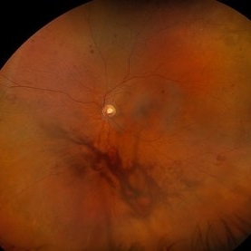
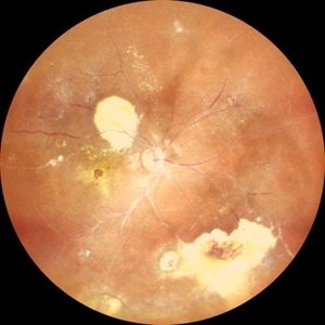 Coats Disease
Sep 29 2024 by Tejaswita Verma
Fundus photo of the RE of a 14 y/o female ,nil premorbid presented with reduced vision in the RE ,diagnosed incidentally on ophthalmological examination elsewhere .Vision was finger counting 3 meters in the RE . Fundus picture reveals macular scar , subretinal and intraretinal exudation ,with scattered hemorrhages esp. in STQ, sclerosed vessels in superior, superonasal quadrant ,nasal, inferonasal quadrant, CR scars inferiorly, Telengiectatic vessels S/O Coat's disease. She was advised RE anti VEGF x1 + laser PRP + PST kenacort under GA with guarded prognosis.
Photographer: DR. TEJASWITA VERMA
Imaging device: MIRANTE
Condition/keywords: Coats' disease
-
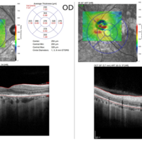 Pattern dystrophies – A 44-year-old man presented with PRPH2 (p.Gln239Ter)-related PD
Sep 17 2024 by Nicolas A Yannuzzi, MD
He had a significant family history of dominant macular dystrophy. OCT images demonstrate intraretinal fluid and disruption of the retinal layers in both eyes. Additionally, vitelliform material can be seen in the right eye and an area of choroidal protrusion in the left. (Images courtesy of Byron L. Lam, MD)
Condition/keywords: inherited retinal disease, pattern dystrophy
-
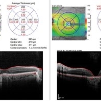 Pattern dystrophies – In a 65-year-old woman with PD, OCT shows bilateral macular atrophy (left worse than right eye) and significant loss of RPE and Bruch membrane
Sep 17 2024 by Nicolas A Yannuzzi, MD
Visual acuity was 20/20 OD and 20/60 OS. Genetics testing showed multiple variants of unknown significance in PEX1, PRPH2, TTLL5, and WFS1. (Images courtesy of Byron L. Lam, MD)
Condition/keywords: inherited retinal disease, pattern dystrophy
 Loading…