 Initializing download.
-
 By Niloofar Piri, MD
SSM Health Group, St Louis University
Co-author(s): Sean Kelso, Saint Louis University - Uploaded on Nov 29, 2022.
- Last modified by Joshua Friedman on Nov 29, 2022.
- Rating
- Appears in
- Miscellaneous
- Condition/keywords
- Rod cone dystrophy, hereditary retinal dystrophy, hereditary retinal deg
- Photographer
- Sean Kelso, Saint Louis University
- Imaging device
- Fundus camera
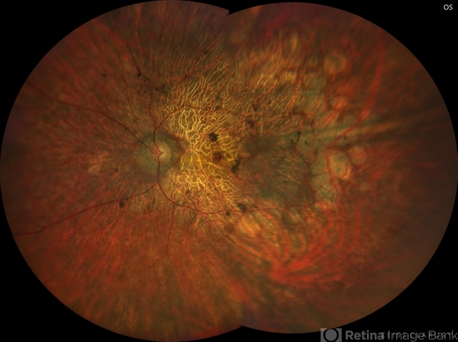
- Description
- Fundus photograph of the left eye in a 58 yo male with rod cone dystrophy. He presented with night blindness and peripheral vision loss since youth and recent decrease in central vision for the past 10 years. Notice waxy pallor of the nerve, severe arterial narrowing and chorioretinal atrophy mainly around the arcades as well as posterior pole along with RPE hyperplastic changes and atrophy. RPE atrophy in midperiphery has coin shaped appearance. FAF has characteristic appearance (uploaded separately) He has one pathogenic variants of both CEP290 and PRPH2 genes.


