Search results (176 results)
-
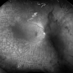
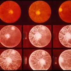
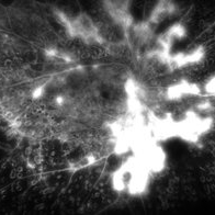
 LASER PRP: LE
Jan 8 2024 by ANKIT JAIN
LEFT EYE WIDEFILED FUNDUS PHOTO OF 38 YEARS OLD FEMALE WITH TYPE 1 DIABETES MELLITUS HAVING WITH 360 DEGREE LASER PRP
Photographer: DR ANKIT JAIN
Imaging device: MIRANTE
Condition/keywords: Diabetes, pan-retinal photocoagulation (PRP), PRP
-
 Proliferative diabetic retinopathy
Aug 22 2023 by rahul saradge
39 year old with unstable PDR s/p PRP , vitreous hemorrhage.
Photographer: Aniket Pednekar , Isha Netralaya
Condition/keywords: PRP, red-free, vitreous blood
-
 Proliferative Diabetic Retinopathy
Aug 11 2025 by Marin Shehata
Fundus photograph of a 63 year-old male with diabetic retinopathy has been treated with PRP.
Photographer: Marin Shehata, Retina Consultants of Carolina
Imaging device: Optos California
Condition/keywords: proliferative diabetic retinopathy (PDR), PRP
-
 Proliferative Diabetic Retinopathy S/P Pan Retinal Photocoagulation
Mar 4 2025 by Prithvi Chandrakanth
A 52-year-old female patient presented with complaints of diminishing vision, compounded by uncontrolled diabetes mellitus. Her Fundus examination revealed proliferative diabetic retinopathy, characterized by neovascularization of the disc and elsewhere, and sclerosed vessels. To address this, Pan Retinal Photocoagulation was performed, and the condition stabilized, halting the progression of the disease.
Photographer: DR PRITHVI CHANDRAKANTH, DR CHANDRAKANTH NETHRALAYA, KOZHIKODE, KERALA, INDIA
Imaging device: EIDON
Condition/keywords: Diabetic Retinopathy, Neovascularisation at the Disc (NVD), neovascularization of the disc (NVD), NVD, pan-retinal photocoagulation (PRP), PDR, PDR with NVE (periphery), PRP
-
 Sickle-Cell Retinopathy
Jan 22 2025 by Virginia Gebhart
Fluorescein angiogram of 54 year old female with non-diabetic proliferative retinopathy. Recent labs confirm sickle-cell disease. FA shows temporal peripheral non perfusion with NV. S/p PRP with retrobulbar block
Photographer: Virginia Gebhart, Retina Consultants of Carolina
Imaging device: Optos California
Condition/keywords: FA, Fluorescein angiography, Neovascularisation elsewhere (NVE), non-perfusion, Nose, pan-retinal photocoagulation (PRP), PRP, sickle cell retinopathy
-
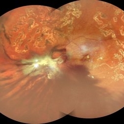 Table Top Tractional Retinal Detachment With Vitreous Hemorrhage in a Case of Proliferative Diabetic Retinopathy
Sep 12 2025 by Akansha Sharma
Color fundus photograph of a 56 year old male with table top tractional retinal detachment with vitreous hemorrhage in a case of proliferative diabetic retinopathy.
Photographer: DR. AKANSHA SHARMA
Condition/keywords: pan-retinal photocoagulation (PRP), PDR, proliferative diabetic retinopathy (PDR), PRP, TABLE TOP TRD, tractional retinal detachment, TRD, VH, vitreous hemorrhage
-
Vitrectomy for Subhyaloid blood over macula in Diabetic retinopathy
Nov 29 2022 by Manish Nagpal, MD, FRCS (UK), FASRS
Subhyaloid blood over macula in diabetic retinopathy| This is a case of non resolving subhyaloid haemorrhage over macula in a case of diabetic retinopathy.. Vitrectomy is carried out and then using the cutter a opening is made in the hyaloid to give a oulet to the blood. Typically the blood in the subhyaloid plane does not clot and easily aspirates out|. After this endolaser PRP is carried out to achieve good regression of the retinopathy. Air fluid exchange is carried out.
Photographer: Manish Nagpal
Condition/keywords: diabetic macular oedema, diabetic retinopathy, endolaser, PRP, SHH, subhyaloid blood, subhyaloid haemorrhage, video, vitrectomy
-
Vitrectomy TRD in Proliferative diabetic retinopathy
Jan 2 2023 by Manish Nagpal, MD, FRCS (UK), FASRS
Vitrectomy for PDR and TRD using Cutter based dissection| This is a case of subhyaloid hemorrhage and Tractional retinal detachment in a diabetic patient. The subhyaloid hemorrhage is aspirated using the cutter . 25 gauge bevelled cutter is used to dissect all the epiretinal proliferations and tractional components. The ports of these cutters can reach very close to the retinal surface and cut flush without causing any iatrogenic damage to the retinal surface. Forceps are used to peel adherent membranes Bleeders are stopped raising pressure and applying diathermy. Once the retina is flattened endolaser is done 360 degree to achieve long term regression.
Condition/keywords: cutter, diabetic retinopathy, endolaser, forceps, PDR, peeling, proliferative diabetic retinopathy (PDR), PRP, tractional retinal detachment, TRD, video, vitrectomy
-
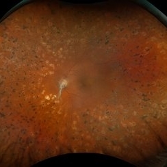
 VITREOUS HEMORRHAGE WITH 360 DEGREE LASER PRP: RE
Jan 8 2024 by ANKIT JAIN
RIGHT EYE WIDEFILED FUNDUS PHOTO OF 38 YEARS OLD FEMALE WITH TYPE 1 DIABETES MELLITUS HAVING VITREOUS HEMORRHAGE WITH 360 DEGREE LASER PRP
Photographer: Dr Ankit Jain
Imaging device: MIRANTE
Condition/keywords: Diabetes, pan-retinal photocoagulation (PRP), PRP, vitreous hemorrhage
-
 PDR
PDR
Jan 29 2014 by Howard Schatz, MD
PRP and CRAO
-
 PRP Day Of Treatment
Oct 8 2012 by Jeffrey G. Gross, MD, FASRS
PRP, day of treatment.
Condition/keywords: pan-retinal photocoagulation (PRP), scatter laser treatment
-
 PRP laser
Mar 29 2013 by Henry J. Kaplan, MD
Right after PRP laser in PDR.
Condition/keywords: laser photocoagulation, pan-retinal photocoagulation (PRP)
-
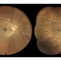
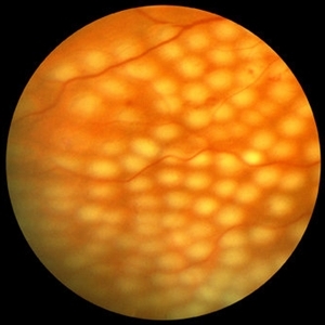
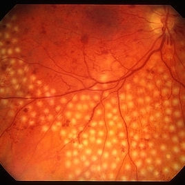
 PRP Marks
Apr 26 2021 by Priya Rasipuram Chandrasekaran, MBBS, DO, DNB, FRCS
This is the fundus photo montage of both eyes of a patient showing pan retinal photocoagulation marks. Theses marks can be confused with gyrate atrophy, cobble stone degeneration and myopic degeneration.
Condition/keywords: pan-retinal photocoagulation (PRP)
-
 28-Year-Old Male With Susac's Syndrome
Feb 2 2015 by Gregory J. Mincey, MD, MBA
Recurrent proliferative disease after initial stabilization with PRP.
Photographer: Bill McVerry, Carolina Eye Associates
Imaging device: Topcon
Condition/keywords: Susac's syndrome
-
 Active diabetic retinopathy despite PRP
Oct 30 2022 by Diego Andrés Rodriguez, MD
A 52-year-old patient with active proliferative diabetic retinopathy despite good glycemic control and PRP performed 1 year ago in the right eye
Photographer: Sociedad de Cirugía Ocular
Imaging device: Clarus 700
Condition/keywords: diabetic retinopathy, pan-retinal photocoagulation (PRP), proliferative diabetic retinopathy (PDR), wide angle imaging
-
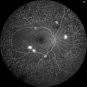
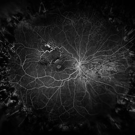
 Active neovascularization in Proliferative Diabetic Retinopathy
Jan 10 2018 by Peter H. Tang, MD, PhD
Fluorescein angiography image from a 46-year-old woman with uncontrolled proliferative diabetic retinopathy shows extensive dye leakage from active neovascularization.
Imaging device: Optos California
Condition/keywords: diabetes, diabetic retinopathy, fluorescein leakage, neovascularization elsewhere (NVE), neovascularization of the disc (NVD), pan-retinal photocoagulation (PRP), proliferative diabetic retinopathy (PDR)
-
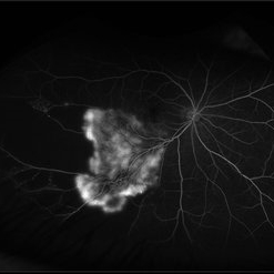
 Active Proliferative Diabetic Retinopathy
Jul 12 2024 by Korey Starkey
Fluorescein angiogram performed on 35 year old female with active proliferative diabetic retinopathy. Patient has peripapillary vascular loop and history of PRP treatment in both eyes. Patients left eye vision measured at Dcc20/200-1 at this visit.
Photographer: Korey Starkey
Imaging device: Optos
Condition/keywords: FLUORESCEIN ANGIOGRAPHY, hyperfluorescence, laser scarring, Optos, proliferative diabetic retinopathy (PDR), sea fan, ultra-wide field imaging, vascular loop
-
 Background Diabetic Retinopathy (BDR)
Sep 5 2013 by Howard Schatz, MD
Right eye 20/50 - DM, PRP.
-
 Brach Retinal Artery Occlusion
Oct 2 2013 by Jerald A. Bovino, MD
There is a hollenhorst plaque causing a branch retinal artery occlusion. The patient has scars from prior panretinal laser photocoagulation.
Condition/keywords: branch retinal artery occlusion (BRAO), hollenhorst plaque, pan-retinal photocoagulation (PRP)
-
 Branch Retinal Artery Occlusion
Oct 2 2013 by Jerald A. Bovino, MD
There is a hollenhorst plaque causing a branch retinal artery occlusion. The patient has scars from prior panretinal laser photocoagulation.
Condition/keywords: branch retinal artery occlusion (BRAO), hollenhorst plaque, pan-retinal photocoagulation (PRP)
-
 Branch Retinal Vein Occlusion
Dec 9 2020 by Olivia Rainey
Ultra-widefield angiogram of a 78-year-old male with a branch retinal vein occlusion affecting his right eye. The patient was diagnosed on 12/17/12 at another practice. The physician noted that there wasn't NVE noted, however areas of micoaneurysmal dilation is present. She noted retinal ischemia secondary to BRVO. 12/8/20 leakage on FA noted to be worsening compared to his previous angiography. She has concern for progressing NVE and recommends sector PRP after injection of antiVEGF series of 3 for the health of the eye.
Photographer: Olivia Rainey, OCT-C, COA
Imaging device: Optos California
Condition/keywords: branch retinal vein occlusion (BRVO), macular branch retinal vein occlusion (BRVO), non-perfusion, scleral buckle, vitreoretinal surgery
-
 Branch Retinal Vein Occlusion with Retinal Neovascularization
Mar 21 2024 by Isaac Agranoff
Fundus angiography photograph of a 63 year old male presenting with worsening blurry vision OD for 4 years with new transient floaters (vision 20/160 PH 20/60). Fluorescein angiography revealed significant capillary non-perfusion corresponding to the area, with peripheral vascular remodeling. Physician recommended anti-VEGF therapy and FA-guided supplemental PRP given the size of the NVE.
Photographer: Isaac Agranoff
Imaging device: Optos California
Condition/keywords: branch retinal vein occlusion (BRVO), EYLEA, FLUORESCEIN ANGIOGRAPHY, Neovascularisation elsewhere (NVE), Optos
-
 BRVO With PRP laser
Feb 19 2015 by H. Michael Lambert, MD
Color photo of NVE after BRVO. Laser performed,ERM present
Condition/keywords: ischemia, neovascularization elsewhere (NVE)
-
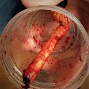 Carotid Artery Plaque
Sep 9 2020 by John S. King, MD
66-year-old white male, former smoker, with a history of femoral artery stent a plaque removal in 2017, triple bypass 2019 (at that time there was no high grade carotid stenosis), diabetes significant for SNPDR OD and PDR OS (NVD). He underwent PRP OS and two months later developed a vitreous hemorrhage and had a PPV OS. Early in the post-operative period vision dropped to LP due to acute CRAO with retinal embolus present. He was found to have progressed to high grade carotid stenosis (versus imaging 6 months ago) and a left carotid endarterectomy was performed (see picture of the large plaque) .
Condition/keywords: carotid artery occlusion
-
 Central Areloar Choroidal Dystrophy
Oct 1 2019 by Demetrios G. Vavvas, MD, PhD
AF of a 31-year-old with CACD type 2 PRPH2 mutation.
Condition/keywords: central areolar choroidal dystrophy (CACD)
 Loading…