Search results (176 results)
-
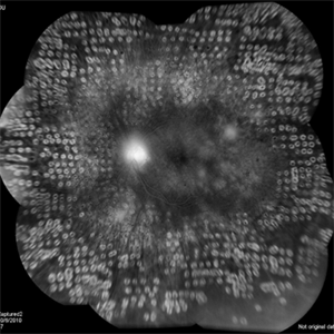
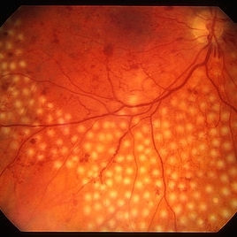 PRP laser
Mar 29 2013 by Henry J. Kaplan, MD
Right after PRP laser in PDR.
Condition/keywords: laser photocoagulation, pan-retinal photocoagulation (PRP)
-
 Regressed Proliferative Diabetic Retinopathy following PRP
Sep 6 2012 by Sharon Fekrat, MD FACS FASRS
58-year-old man with regressed proliferative diabetic retinopathy in the left eye following panretinal laser photocoagulation. Note attenuated retinal vasculature.
Photographer: Sarah Enfiedjian, Ophthalmic Photographer, Durham VA Medical Center, Durham, NC
Imaging device: Zeiss
Condition/keywords: attenuated vessels, pan-retinal photocoagulation (PRP)
-
 PDR with Active NVD
Oct 8 2012 by Jeffrey G. Gross, MD, FASRS
PDR with active NVD and preretinal hemorrhage, mild VH and partial PRP.
Condition/keywords: neovascularization of the disc (NVD), preretinal hemorrhage, scatter laser photocoagulation, vitreous hemorrhage
-
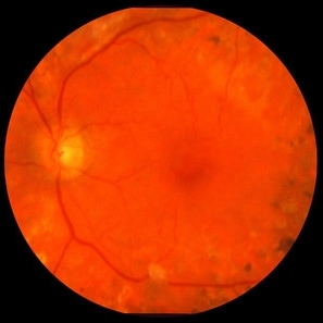
 Diabetic Retinopathy
Oct 18 2012 by Raj K. Maturi, MD
Photographer: Tom Steele, CRA
Imaging device: Optos
Condition/keywords: pan-retinal photocoagulation (PRP)
-
 PRP Day Of Treatment
Oct 8 2012 by Jeffrey G. Gross, MD, FASRS
PRP, day of treatment.
Condition/keywords: pan-retinal photocoagulation (PRP), scatter laser treatment
-
---thumb.jpg/image-square;max$300,300.ImageHandler) Proliferative Diabetic Retinopathy (PDR), PRP, Regressed NV
Proliferative Diabetic Retinopathy (PDR), PRP, Regressed NV
Feb 13 2013 by From the Collections of Thomas M. Aaberg, MD and Thomas M. Aaberg Jr., MD
5 months s/p, PRP, Pt. GL.
Condition/keywords: neovascularization (NV), pan-retinal photocoagulation (PRP)
-
---thumb.jpg/image-square;max$300,300.ImageHandler) Proliferative Diabetic Retinopathy (PDR), PRP, Regressed NV
Proliferative Diabetic Retinopathy (PDR), PRP, Regressed NV
Feb 13 2013 by From the Collections of Thomas M. Aaberg, MD and Thomas M. Aaberg Jr., MD
1 year, s/p, PRP.
Condition/keywords: neovascularization (NV), pan-retinal photocoagulation (PRP), post-laser
-
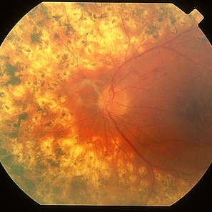 Regressed PDR
Mar 29 2013 by Henry J. Kaplan, MD
Regressed PDR after full PRP, notice the FPD on the optic nerve.
Condition/keywords: pan-retinal photocoagulation (PRP), regressed
-
 Marked Retinal Ischemia in Patient with Mixed Connective Tissue Disease
Feb 26 2013 by Sharon Fekrat, MD FACS FASRS
Fluorescein angiogram of the right eye of a 27-year-old female with mixed connective tissue disease and marked retinal ischemia. Panretinal laser photocoagulation (PRP) has been performed for neovascularization elsewhere (NVE).
Condition/keywords: mixed connective tissue disease, retinal ischemia
-
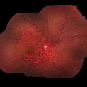
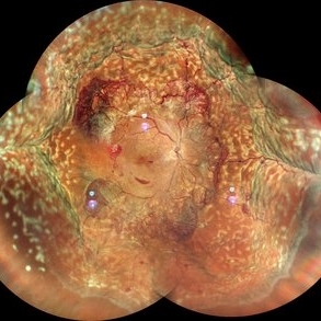 Proliferative Diabetic Retinopathy with Choroidal Effusion Status Post PRP
Dec 15 2020 by Manish Nagpal, MD, FRCS (UK), FASRS
A 17-year-old juvenile diabetic patient came to us with extensive neovascular proliferations and PRP done a week back and had developed 360 degree choroidal effusion as seen in this wide field montage image
Photographer: Sham Talati, Retina Fellow , Retina Foundation, Ahmedabad, India
Imaging device: Mirante CSLO
Condition/keywords: choroidal effusion, diabetic retinopathy, proliferative diabetic retinopathy (PDR)
-
 Wyburn Mason Racemose Angiomatosis
May 22 2016 by Olivia Rainey
Color fundus montage of an 13-year-old female with arteriovenous malformation (Wyburn Mason Racemose Angiomatosis) affecting her right eye. The retinal arteriovenous malformation appears to be stable. She presented with NLP in the eye, strabismus, and peripheral retinal ischemia. She is at risk for neovascular complications; however, she is currently being treated with Sirolimus. Since she is on this systemically, there is no need to perform intraocular anti-VEGF injections or PRP laser. She also presented with optic atrophy affecting her left eye, secondary to chiasmal involvement of arteriovenous malformation. She has had a potential progressive visual field loss involving the temporal aspect of her visual field from the left eye. There is sector optic atrophy. Presumably, this is due to a compressive effect of her arteriovenous malformation on the nasal nerve fiber layer (corresponding to the temporal visual field) crossing to the right occipital cortex at the chiasm.
Photographer: Olivia Rainey
Imaging device: Topcon 50dx
Condition/keywords: arteriovenous malformation, color fundus photograph, color photo, montage, peripheral ischemia, Sirolimus
-
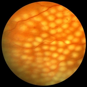
 Extensive Pan-Retinal Photocoagulation
Apr 19 2013 by Brandon G. Busbee, MD
Extensive pan-retinal photocoagulation.
Photographer: Alecia Camp, CRA - Tennessee Retina - Nashville, TN
Imaging device: Topcon TRC 50-EX
Condition/keywords: neovascularization (NV), pan-retinal photocoagulation (PRP)
-
 Proliferative Diabetic Retinopathy with Macular Traction
Oct 15 2012 by Jeffrey G. Gross, MD, FASRS
PDR with macular traction, post-op, PPV and PRP.
Condition/keywords: macular traction, pan-retinal photocoagulation (PRP), pars plana vitrectomy (PPV), post-op
-
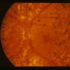
 Diabetic Retinopathy
Oct 18 2012 by Raj K. Maturi, MD
Photographer: Tom Steele, CRA
Imaging device: Optos
Condition/keywords: pan-retinal photocoagulation (PRP)
-
 Branch Retinal Artery Occlusion
Oct 2 2013 by Jerald A. Bovino, MD
There is a hollenhorst plaque causing a branch retinal artery occlusion. The patient has scars from prior panretinal laser photocoagulation.
Condition/keywords: branch retinal artery occlusion (BRAO), hollenhorst plaque, pan-retinal photocoagulation (PRP)
-
---thumb.jpg/image-square;max$300,300.ImageHandler) Proliferative Diabetic Retinopathy (PDR), PRP, Regressed NV
Proliferative Diabetic Retinopathy (PDR), PRP, Regressed NV
Feb 13 2013 by From the Collections of Thomas M. Aaberg, MD and Thomas M. Aaberg Jr., MD
5 months s/p. PRP, Pt. GL.
Condition/keywords: neovascularization (NV), pan-retinal photocoagulation (PRP)
-
 Brach Retinal Artery Occlusion
Oct 2 2013 by Jerald A. Bovino, MD
There is a hollenhorst plaque causing a branch retinal artery occlusion. The patient has scars from prior panretinal laser photocoagulation.
Condition/keywords: branch retinal artery occlusion (BRAO), hollenhorst plaque, pan-retinal photocoagulation (PRP)
-
 Diabetic Retinopathy
Oct 18 2012 by Raj K. Maturi, MD
Photographer: Tom Steele, CRA
Imaging device: Optos
Condition/keywords: pan-retinal photocoagulation (PRP)
-
 Exudative Macular Detachment After Intensive Laser Photocoagulation
Mar 12 2016 by Sjakon G Tahija, MD
Fundus photograph of 44-year-old man with exudative detachment of the macula after vitrectomy and ILM peeling for proliferative diabetic retinopathy combined with intensive endolaser photocagulation.
Photographer: Avris Siahaan, Klinik Mata Nusantara
Condition/keywords: exudative detachment, pan-retinal photocoagulation (PRP)
-
 persisting NVD after heavy PRP
Jan 1 2013 by John T. Thompson, MD
Persisting severe NVD despite heavy PRP for PDR.
Condition/keywords: neovascularization of the disc (NVD)
-
 Neovascularization of the Disc
Apr 19 2013 by Brandon G. Busbee, MD
Persistant NVD after extensive PRP.
Photographer: Alecia Camp, CRA - Tennessee Retina - Nashville, TN
Imaging device: Topcon TRC 50-EX
Condition/keywords: neovascularization of the disc (NVD), pan-retinal photocoagulation (PRP)
-
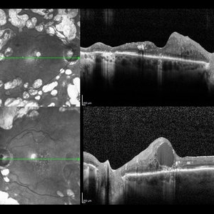 Diabetic Macular Edema
May 28 2016 by Olivia Rainey
Optical coherence tomography of an 54-year-old female with diabetic macular edema affecting both eyes. Patient has a history of proliferative diabetic retinopathy s/p PRP/PPV/MP/EL, and glaucoma s/p tube shunt in both eyes. There has been a persistence of her macular edema and limited response to antiVEGF therapy, which puts into question whether there is another cause for her edema. Leading the possible causes is her renal insufficiency and fluid retention. Patient was seeing 20/50 in the right eye and 20/80 in the left eye.
Photographer: Olivia Rainey
Imaging device: Heidelberg Spectralis
Condition/keywords: anti-VEGF, diabetic macular edema, edema, glaucoma, optical coherence tomography (OCT), pan-retinal photocoagulation (PRP), proliferative diabetic retinopathy (PDR)
-
 Proliferative Diabetic Retinopathy with PRP
Mar 9 2013 by Gabriela Lopezcarasa Hernandez, MD
Proliferative diabetic retinopathy with PRP.
Photographer: Azucena Rios, Macula Retina Consultores Mexico
Imaging device: Heidelberg Spectralis
Condition/keywords: argon photocoagulation
-
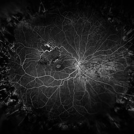 Branch Retinal Vein Occlusion
Dec 9 2020 by Olivia Rainey
Ultra-widefield angiogram of a 78-year-old male with a branch retinal vein occlusion affecting his right eye. The patient was diagnosed on 12/17/12 at another practice. The physician noted that there wasn't NVE noted, however areas of micoaneurysmal dilation is present. She noted retinal ischemia secondary to BRVO. 12/8/20 leakage on FA noted to be worsening compared to his previous angiography. She has concern for progressing NVE and recommends sector PRP after injection of antiVEGF series of 3 for the health of the eye.
Photographer: Olivia Rainey, OCT-C, COA
Imaging device: Optos California
Condition/keywords: branch retinal vein occlusion (BRVO), macular branch retinal vein occlusion (BRVO), non-perfusion, scleral buckle, vitreoretinal surgery
-
 Diabetic Retinopathy
Aug 21 2015 by Andrea Arriola-Lopez, MD MSc
Color fundus photography shows neovascularization of the optic nerve head, macular pre retinal hemorrhage, pan retinal photocoagulation and extreme temporal peripherical retina without PRP.
Photographer: Andrea Elizabeth Arriola L.
Imaging device: OPTOS Dakota
Condition/keywords: diabetes, diabetic retinopathy, neovascularization (NV)
 Loading…