Search results (171 results)
-
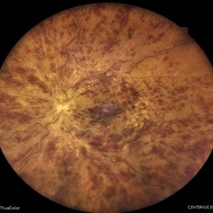
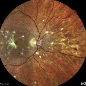
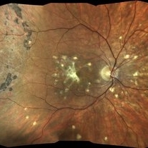
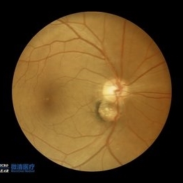
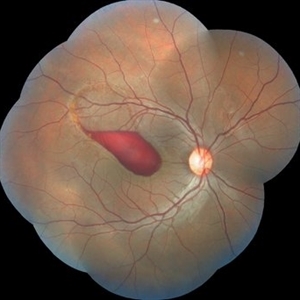
 Myelinated Nerve Fibers
Apr 18 2025 by DR Rohit Gupta
The **myelinated nerve fibers of the optic disc** (also known as **medullated nerve fibers**) are retinal nerve fibers that retain their myelin sheath as they pass through the optic nerve head. Normally, retinal nerve fibers are unmyelinated to allow for light transparency, but in some cases, myelination extends anteriorly into the retina, appearing as a striking white, feathery patch on the optic disc or peripapillary retina. ### **Key Features:** 1. **Appearance:** - Dense, white, striated patches with feathery edges. - Typically located at the superior or inferior pole of the optic disc. - May obscure retinal vessels underneath. 2. **Clinical Significance:** - Usually **benign** and asymptomatic. - **Congenital** (present at birth or early childhood). - Rarely associated with **visual field defects** (e.g., scotomas corresponding to the area of myelination). - Occasionally linked with **high myopia** or **amblyopia** if extensive. 3. **Pathophysiology:** - Failure of oligodendrocytes or Schwann cells to stop myelination at the lamina cribrosa. - Normally, myelination stops at the optic nerve head, but in this condition, it extends into the retina. 4. **Diagnosis:** - **Fundoscopy:** Classic white, feathery appearance. - **Optical Coherence Tomography (OCT):** Shows thickened retinal nerve fiber layer (RNFL). - **Visual Field Testing:** May detect defects if large. 5. **Differential Diagnosis:** - Optic disc edema - Cotton wool spots - Retinoblastoma (rarely, but must be ruled out in children) 6. **Management:** - No treatment required if asymptomatic. - Monitor for amblyopia in children. - Rare cases with significant visual impairment may need further evaluation. ### **Fun Fact:** Myelinated nerve fibers are seen in **~0.5-1%** of the population and are usually an incidental finding.
Photographer: Dr Rohit gupta
Imaging device: Samsung S21
Condition/keywords: Medulated Nerve fibre, Medullated Nerve fibres, myelinated nerve fibers, Myelinated Nerve Fibres, optic disc drusen
-
 Central Retinal Vein Occlusion
Feb 25 2025 by Prithvi Chandrakanth
A 61-year-old woman with a history of hypertension noticed a sudden painless blurring of vision in her left eye. Over the next few days, the blurriness persisted, and she experienced a mild central scotoma. On examination, Fundoscopic evaluation revealed dilated, tortuous retinal veins, retinal hemorrhages, and macular oedema.
Photographer: DR.PRITHVI CHANDRAKANTH, DR.CHANDRAKANTH NETHRALAYA, KOZHIKODE
Imaging device: EIDON
Condition/keywords: CRVO, CRVO WITH MACULA EDEMA, flame shaped retinal hemorrhage
-
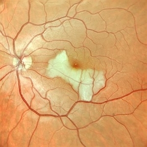
 Myopic CNVM
Jan 31 2025 by Thirumalesh Mochi Basavaraj, MD
Widefield image of a 26 year-old male patient with pathologic myopia with history of central scotoma with a sub macular bleed.
Photographer: Puttaswamy N K
Imaging device: Optos Daytona
Condition/keywords: myopic choroidal neovascularization (CNV), Myopic CNVM, pathologic myopia
-
 Subhyaloid Hemorrhage
Jul 31 2024 by Arthi Mohankumar , MS,MRCS ED, FICO,FAICO
A 35 year old male presented with complaints of seeing a black spot in left eye for past one day after working out in the gym the previous day. He has history of uncontrolled diabetes and hypertension. Fundus exam of the left eye revealed a sub hyaloid hemorrhage nasal to the disc with minimal background Diabetic and hypertensive changes. His baseline CBG was 200 mg/dl and BP was 170/100 He was suggested observation initially considering the nasal location. But patient found the scotoma very disturbing and eventually underwent yag hyaloidotomy
Photographer: Arthi Mohankumar
Condition/keywords: Sub hyaloid haemorrhage, valsalva retinopathy
-
 Cilioretinal Artery Occlusion
May 14 2024 by Eloy Mata-Cortes, MD
Color image capturing the left eye of a 32-year-old female. Despite a negative ophthalmological and medical history, she reported three days of blurred vision and a paracentral scotoma in her left eye, while maintaining central vision. The image reveals retinal whitening, extends from the parafoveal region to the inferotemporal arcade indicative of cilioretinal artery occlusion. Following this observation, the patient was referred for systemic assessment to explore the underlying etiology of the occlusion.
Photographer: Eloy Mata-Cortes, MD, Instituto Mexicano de Oftalmología, Querétaro, México
Imaging device: Nidek Mirante
Condition/keywords: cilioretinal artery occlusion, oclussion, retinal whitening
-
 Best Disease
Apr 24 2024 by Marcelo Zas, MD PhD
Best vitelliform macular dystrophy (BVMD) or Best disease. Is the most common autosomal dominant macular dystrophy. It involves the retinal pigment epithelium (RPE), and leads to a characteristic bilateral yellow “egg-yolk” appearance of the macula as you can see in this image. Essentially, BVMD is considered to have 6 clinical stages: Previtelliform, Vitelliform, Pseudohypopyon, Vitelleruptive, Atrophic and Choroidal neovascularization. As the disease progresses, patients may experience a slow, bilateral decrease in visual acuity, central scotoma, or metamorphopsia. With secondary CNV, visual decline can be rapid, however.
Photographer: Luciano Scorsetti MD
Condition/keywords: Macular Dystrophy
-
 Left Eye Arteriovenous Malformation, Vein Occlusion and Ruptured Macroaneurysm
Feb 9 2024 by Sandra R Montezuma, MD
47 year old female presented with acute changes in vision in the left eye, flashes of light and a new supero temporal scotoma. No history of trauma. She has history of retina bleeding in 1998 when she was pregnant and had pre-eclampsia. She was told had a retina scar. Her VA was 20/500. Fundus exam revealed an arteriovenous malformation along inferonasal vessels with prominent tortuous vessels. The optic nerve was hyperemic and there was peripapillary pre-retinal hemorrhage. There is a central macula scar and retina hemorrhage in the macula and mid periphery. In the nasal mid periphery, there is a ruptured macroaneurysm with hemorrhage in all layers of the retina. There are diffuse IRH. Her OCT revealed abnormal foveal contour with intraretinal fluid, Outer retinal atrophy and increased hyperreflectivity of the inner retina layers. The patient was treated with avastin injections with some improvement of the vision and resolution of the intraretinal fluid. Her MRI was normal.
Photographer: University of Minnnesota
Condition/keywords: arteriovenous malformation, macroaneurysm, vein occlusion
-
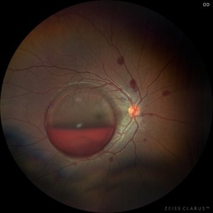
 Retinal Detachment
Jan 31 2024 by Cuitláhuac del Moral Herrera
69 year old male presented with a week long scotoma on his right eye. A macula ON retinal detachment with a “spokes-wheel” giant tear was noted upon examination. We performed a phaco-vit with good results.
Photographer: Manuela Franco Sánchez, Asociación para Evitar la Ceguera en México. Cuitláhuac del Moral Herrera, Asociación para Evitar la Ceguera en México.
Imaging device: ZEISS CLARUS 700
Condition/keywords: macula on, Spokes wheel
-
 Left Inferior Quadrantopia - Microperimetry
Jan 17 2024 by Francisco Fraga Santini Canto
Microperimetry of a 63-year-old male with sequelae of ischemic stroke in the right parietal lobe years ago. In the right eye, the patient also had a central scotoma secondary to silicone oil toxicity.
Photographer: Leonardo Hideki Nomachi Naito
Imaging device: Navis-EX Microperimeter
Condition/keywords: ischemic stroke, microperimeter, neuro-ophtalmolgy, silicone oil toxicity, visual field defect
-
 Punctate inner choroidopathy (PIC) with CNVM
Oct 18 2023 by Heitor Nogueira
Fundus photograph of a 29-year-old woman with a 2-week history of low visual acuity associated with central scotoma. Ophthalmological history of axial myopia of -4,00D. She denied personal and family history.
Photographer: Heitor Nogueira, Instituto Penido Burnier, Campinas-SP, Brazil.
Imaging device: Eidon True Color
Condition/keywords: CNVM, multifocal chorioretinitis (MCP), punctate inner choroidopathy (PIC)
-
 Punctate inner choroidopathy (PIC) with CNVM.
Oct 18 2023 by Heitor Nogueira
Fundus photograph of a 29-year-old woman with a 2-week history of low visual acuity associated with central scotoma. Ophthalmological history of axial myopia of -4,00D. She denied personal and family history.
Photographer: Heitor Nogueira, Instituto Penido Burnier, Campinas-SP, Brazil.
Imaging device: Eidon True Color
Condition/keywords: choroidal neovascularization (CNV), CNVM, multifocal chorioretinitis (MCP), punctate inner choroidopathy (PIC)
-
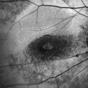 Plaquenil-toxicity
Mar 31 2023 by Niloofar Piri, MD
Fundus autofluorescence image of the right eye in a highly myopic Caucasian patient who was screened for plaquenil toxicity elsewhere for 15 years. Medicine was stopped 3 years ago. She presented to us with deteriorationg central vision and scotoma for the past 3 years. FAF demonstrtaes classic symmetric bull's eye pattern of hypoautofluorescence in parafoveal area both eyes with some extension to the arcades, which is the result of severe plaquenil toxicity. Notice the hyperAF area surrounding the bull'e eye which is demonstarting stressed RPE cells filled with Lipofuscin. This area will likely turn to a larger hypoAF later. It is critical to diagnose it early to prevent the ongoing damage and loss of RPE and photoreceptors from the long term deposited medicine in RPE cells.
Photographer: Sean Kelso, Saint Lousi University
Condition/keywords: bull's eye atrophy, bull's eye maculopathy, hydroxychloroquine toxicity, plaquenil toxicity
-
 Valsalva Retinopathy
Nov 18 2022 by Niloofar Piri, MD
21 yo female presented with decaresed central vision and scotoma immediately after severe vomiting. Color fundus phtograph demonstrates large sub ILM layered hemorrhage in the macula consistent with valsalva retinopathy. Notice the sacttered blot retinal hemorrhages in mid-periphery.
Photographer: Rocio Bentivegna, MD, Saint Louis University
Condition/keywords: sub ILM hemorrhage, valsalva retinopathy
-
 Double disc sign
Oct 13 2022 by Vaibhavi Noticewala, M S Ophthalmology, FVRS
Double disc sign Doubling of the optic disc is rare and can manifest as true or pseudo doubling. Duke-Elder describes duplication of the optic disc as a rare anomaly wherein two discs, each provided with retinal vessels are seen in an otherwise normal eye. Rare cases of true duplication of optic discs with separation of optic nerve into two or more strands have been reported, based either on incidental necropsy findings, demonstration of two optic foramina in the same orbit on x ray, or angioscotomas as indirect evidence of the existence of double optic nerves. Pseudo doubling of the optic discs caused by lesions such as optic disc coloboma, peripapillary chorioretinal coloboma, or inflammatory foci are more common. Our case had Ipsilateral isolated ectatic peripapillary chorioretinal coloboma simulating double optic discs.
Photographer: Priyal Mistry
Condition/keywords: Pseudoduplication of optic disc
-
 Acute syphilitic posterior placoid chorioretinitis
Apr 24 2022 by Aniruddha K Agarwal, MD
Green-light fundus autofluorescence (FAF) of the right eye from a 55-year-old man with risk factors for sexually trasnmitted diseases who presented to the retina clinic for a central scotoma. Funduscopy revealed a placoid lesion in the posterior pole. FAF highlights a hyperautofluorescent placoid lesion involving the macula with granular hyperfluorescence. The patient tested positive for syphilis and received intravenous penicillin treatment.
Photographer: Esther CIANCAS, MD, PhD, Gema CRESPO-RODRÍGUEZ, RN
Imaging device: Zeiss Clarus fundus camera
Condition/keywords: chorioretinitis, IUSG, syphilis, uveitis
-
 Sub-Internal Limiting Membrane Hemorrhage - Pre and Post YAG Laser
May 21 2021 by Anmol Naik
A 36-year-old male complained of central scotoma in the right eye after observing intense laser lights at a local festival celebration. His BCVA was 6/60. On examination, he had a sub-internal limiting membrane (ILM) hemorrhage, which was treated with focal frequency-doubled Nd:YAG laser. Two weeks later, the hemorrhage resolved completely with BCVA of 6/6.
Photographer: Anmol Naik, MS, Insight Institute of Ophthalmology, Pune, India.
Imaging device: Topcon 3D Maestro 1, integrated Fundus camera and OCT
Condition/keywords: focal laser, laser photocoagulation, sub-inner limiting membrane hemorrhage
-
 Choroidal MRSA Abscess
Apr 15 2021 by Rui Zhang, BA
A 14-year-old boy receiving induction chemotherapy for acute lymphocytic leukemia (ALL) complained of floaters and central scotoma in his left eye. (A) Fundus photography showed sub-macular choroidal abscess with intraretinal hemorrhage and edema. (B) OCT confirmed that the abscess had not penetrated the retinal pigment epithelium (RPE). Due to systemic septic signs (fever, tachycardia, tachypnea, new-onset papules), blood cultures were drawn and they came back positive for methicillin-resistant staphylococcus aureus (MRSA). Patient was promptly treated with both IV and intravitreal antibiotics. This is a case of sub-macular choroidal MRSA abscess in the setting of systemic bacteremia in an immunocompromised host.
Photographer: Raymond Mok, CRA COMT (Dartmouth-Hitchcock Medical Center)
Imaging device: Optical coherence tomography
Condition/keywords: abscess, acute leukemia, MRSA sepsis
-
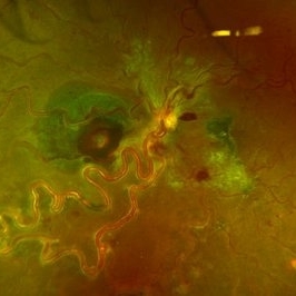
 Acute Macular Neuroretinopathy
Apr 12 2021 by Iuri Golubev, MD
46-year-old female with sudden onset paracentral scotoma below the central point of fixation in her left eye. Enface image shows a wedge shaped lesion pointing towards the fovea (top left). The lesion was spanning outer retinal layers from OPL to RPE (top left insert). One month later, the lesion has diminished in size, and was only involving retinal layers from ellipsoid zone to RPE(top right). At 4 months since presentation, the patient did not have any signs of AMN identifiable on enface or b-scan images (bottom center). Patient's symptoms has slowly improved and eventually resolved over the course of the next 4 years.
Imaging device: Zeiss Cirrus 5000
Condition/keywords: acute macular neuroretinopathy, acute macular outer retinopathy
-
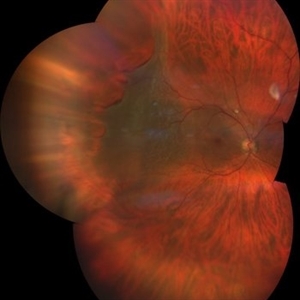
 Rhegmatogenous Retinal Detachment
Mar 3 2021 by Patrik Rajs
A 51-year-old female patient presented with inferior nasal scotoma and 5/10 vision in the right eye due to a retinal detachment with a giant retinal horseshoe tear.
Photographer: Patrik Rajs, EYE CLINIC of Jan Evangelista Purkyne University and Masaryk Hospital, Czech Republic, Ústí nad Labem
Imaging device: Clarus 700
Condition/keywords: giant retinal tear
-
 Multiple Evanescent White Dot Syndrome
Feb 10 2021 by Cláudia Farinha
Ultra-widefield color and autofluorescence of a 30-year-old myopic female with decreased visual acuity, photopsias, and temporal scotomata.
Photographer: Claudia Farinha, MD
Imaging device: Optomap, Optos
Condition/keywords: multiple evanescent white dot syndrome (MEWDS)
-
 Relentless Placoid Chorioretinitis
Jan 22 2021 by Renata Garcia Franco, Md
20-year-old male with reduction of vision in both eyes, scotoma and metamorphopsia. Widespread multiple chorioretinal lesions with RPE hyperplasia, which appear from posterior pole to peripheral retina and inactive choroidal neovascular membrane.
Photographer: Fatima Hernandez, Instituto de la Retina del Bajio SC
Imaging device: Zeiss
Condition/keywords: chorioretinitis
-
 Relentless Placoid Chorioretinitis
Jan 22 2021 by Renata Garcia Franco, Md
20-year-old male with reduction of vision in both eyes, scotoma and metamorphopsia. Widespread multiple chorioretinal lesions with RPE hyperplasia, which appear from posterior pole to peripheral retina.
Photographer: Fatima Hernandez, Instituto de la Retina del Bajio SC
Imaging device: Zeiss
Condition/keywords: chorioretinitis
-
 OCT Showing Premacular Hemorrhage
Nov 26 2020 by Priya Rasipuram Chandrasekaran, MBBS, DO, DNB, FRCS
A 26-year-old male with no h/o trauma or underlying systemic disease presented with the complaint of central scotoma in the right eye since 1 month and fundus examination showed preretinal hemorrhage in the supero-temporal quadrant extending into the macular area and OCT macula showing premacular hemorrhage.
Condition/keywords: premacular hemorrhage
-
 Preretinal Hemorrhage Extending into the Macula
Nov 26 2020 by Priya Rasipuram Chandrasekaran, MBBS, DO, DNB, FRCS
A 26-year-old male with no h/o trauma or underlying systemic disease presented with the complaint of central scotoma in the right eye since 1 month and fundus examination showed preretinal hemorrhage in the supero-temporal quadrant extending into the macular area and OCT macula showing premacular hemorrhage.
Condition/keywords: preretinal hemorrhage
-
 MEWDS
MEWDS
Oct 9 2020 by David L Kilpatrick, MD
26-year-old female presented with unilateral vision loss. She c/o flashes and a peripheral scotoma. Vision was 20/100. On exam, she showed foveal granularity, mild disc edema, and white dots as seen. Three weeks later, white dots had resolved and vision improved to 20/25.
Photographer: Mississippi Retina Associates
Imaging device: Optos
Condition/keywords: multiple evanescent white dot syndrome (MEWDS)
 Loading…