File number: 130309
Comments
-
 Sandra R Montezuma, MD (March 17 2024)
thank you for the comment, do you think is this test better than MRA of the head and neck?
-
 Selma Milisic (March 16 2024)
Maybe to do color doplers of vessels neck and transcranial
Sign in to comment.
 Initializing download.
-
 By Sandra R Montezuma, MD
University of Minnesota
Co-author(s): Adam Neuhouser, MD and Pedro Monsalve, MD University of Minnesota - Uploaded on Feb 9, 2024.
- Last modified by Joshua Friedman on Feb 12, 2024.
- Rating
- Appears in
- 9-Feb-2024
- Condition/keywords
- arteriovenous malformation, macroaneurysm, vein occlusion
- Photographer
- University of Minnnesota
- Imaging device
- Fundus camera
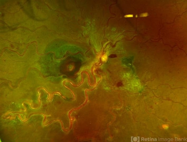
- Description
- 47 year old female presented with acute changes in vision in the left eye, flashes of light and a new supero temporal scotoma. No history of trauma. She has history of retina bleeding in 1998 when she was pregnant and had pre-eclampsia. She was told had a retina scar. Her VA was 20/500. Fundus exam revealed an arteriovenous malformation along inferonasal vessels with prominent tortuous vessels. The optic nerve was hyperemic and there was peripapillary pre-retinal hemorrhage. There is a central macula scar and retina hemorrhage in the macula and mid periphery. In the nasal mid periphery, there is a ruptured macroaneurysm with hemorrhage in all layers of the retina. There are diffuse IRH. Her OCT revealed abnormal foveal contour with intraretinal fluid, Outer retinal atrophy and increased hyperreflectivity of the inner retina layers. The patient was treated with avastin injections with some improvement of the vision and resolution of the intraretinal fluid. Her MRI was normal.


---thumb.jpg/image-square;max$79,0.ImageHandler "Arterial Macroaneurysm")
---thumb.JPG/image-square;max$79,0.ImageHandler "Macroaneurysm")

---thumb.jpg/image-square;max$79,0.ImageHandler "Subretinal Hemorrhage Secondary to Macroaneurysm")
---thumb.jpg/image-square;max$79,0.ImageHandler "Subretinal Hemorrhage Secondary to Macroaneurysm")


