Search results (171 results)
-
 Traumatic Macular Hole
Aug 23 2012 by Gabriela Lopezcarasa Hernandez, MD
12-year-old boy with blunt trauma in right eye and central scotoma.
Photographer: Gabriela Lopezcarasa Hernandez, Hospital Angeles Lomas
Imaging device: ZEISS F4
Condition/keywords: blunt trauma, central scotoma, macular hole
-
 Rhegmatogenous Retinal Detachment
Aug 23 2012 by Gabriela Lopezcarasa Hernandez, MD
30-year-old male with floaters and inferonasal scotoma.
Photographer: Gabriela Lopezcarasa Hernandez, Hospital Angeles Lomas
Imaging device: ZEISS FF4
Condition/keywords: floaters, inferonasal scotoma
-
 Acute Posterior Multifocal Placoid Pigment Epitheliopathy
Sep 15 2012 by Roy D. Brod, MD
Fundus photograph right eye demonstrating cream colored placoid lesions in 28-year-old male patient with 4-day history of multiple scotomas OU.
Photographer: Julia Walker
Condition/keywords: acute posterior multifocal placoid pigment epitheliopathy (APMPPE), placoid retinal lesions, scotoma
-
 Acute Posterior Multifocal Placoid Pigment Epitheliopathy
Sep 15 2012 by Roy D. Brod, MD
Fundus photograph left eye demonstrating cream colored placoid lesions in 28-year-old male patient with 4-day history of multiple scotomas OU.
Photographer: Julia Walker
Condition/keywords: acute posterior multifocal placoid pigment epitheliopathy (APMPPE), placoid retinal lesions, scotoma
-
---thumb.jpg/image-square;max$300,300.ImageHandler) Cilioretinal Artery Occlusion with Central Retinal Vein Occlusion
Cilioretinal Artery Occlusion with Central Retinal Vein Occlusion
Mar 9 2013 by Gabriela Lopezcarasa Hernandez, MD
A 46-year-old male with decrease in visual acuity in left eye and central scotoma.
Photographer: Araceli Rojas Arriaga, Hospital Angeles Lomas, Mexico
Imaging device: Zeiss FF4
Condition/keywords: central retinal vein occlusion (CRVO), cilioretinal artery occlusion
-
 Plaquenil Toxicity
Apr 30 2013 by Theodore Leng, MD, MS, FASRS
Right eye 10-2 HVF from a 44-year-old woman with bilateral plaquenil toxicity. A ring scotoma is present.
Condition/keywords: hydroxychloroquine toxicity, plaquenil toxicity
-
 Myopic Choroidal Neovascularization
Aug 23 2012 by Gabriela Lopezcarasa Hernandez, MD
19-year-old male who complains of scotoma and metamorphopsias.
Photographer: Gabriela Lopezcarasa Hernandez, Macular Retina Consultores
Imaging device: Heidelberg Spectralis
Condition/keywords: choroidal neovascularization (CNV), myopia
-
---thumb.jpg/image-square;max$300,300.ImageHandler) Cilioretinal Artery Occlusion with Central Retinal Vein Occlusion
Cilioretinal Artery Occlusion with Central Retinal Vein Occlusion
Mar 9 2013 by Gabriela Lopezcarasa Hernandez, MD
A 46-year-old male with decrease in visual acuity in left eye and central scotoma.
Photographer: Araceli Rojas Arriaga, Hospital Angeles Lomas, Mexico
Imaging device: Zeiss FF4
Condition/keywords: central retinal artery occlusion (CRAO), central retinal vein occlusion (CRVO), cilioretinal artery occlusion
-
 Plaquenil Toxicity
Apr 30 2013 by Theodore Leng, MD, MS, FASRS
Left Eye 10-2 HVF from a 44-year-old woman with bilateral plaquenil toxicity. A ring scotoma is present.
Condition/keywords: hydroxychloroquine toxicity, plaquenil toxicity
-
 AZOOR
AZOOR
Aug 24 2012 by Geoffrey G. Emerson, MD, PhD, FASRS
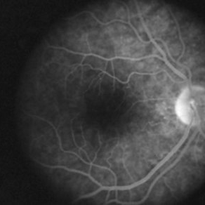
A 17-year-old healthy woman noticed a pacman-shaped scotoma in her temporal right vision. Acuity measured 20/20 and color vision measured 11/11. Angiography showed some late staining of the nasal macula.
Photographer: Geoffrey Emerson, MD, PhD, Retina Center, Minneapolis
Condition/keywords: scotoma
-
 AZOOR
AZOOR
Aug 24 2012 by Geoffrey G. Emerson, MD, PhD, FASRS
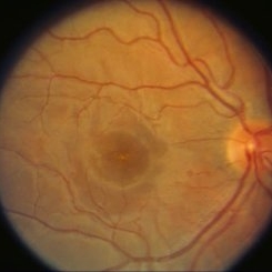
A 17-year-old healthy woman noticed a pacman-shaped scotoma in her temporal right vision. Acuity measured 20/20 and color vision measured 11/11. Possible pigmentary changes were visible at the fovea.
Photographer: Geoffrey Emerson, MD, PhD, Retina Center, Minneapolis
Condition/keywords: scotoma
-
 Valsalva retinopathy mild
Oct 22 2012 by Ronald C. Gentile, MD
A 26-year-old man noticed a central and para-central scotoma in his left eye following strenuous exercise. He was doing push-up while standing on his head. The scotoma and retinal hemorrhage resolved in 2 months with observation.
Photographer: The New York Eye & Ear Infirmary Department of Medical Imaging
Condition/keywords: valsalva retinopathy
-
---thumb.jpg/image-square;max$300,300.ImageHandler) Terson's Syndrome
Terson's Syndrome
Oct 8 2013 by Maurice F. Rabb
39 year female with a long history of chronic back pain treated by a sequence of epidural injections. Following her last injection, she complained of a moderately severe protracted headache and had several attempts at placement of an epidural blood patch without success. Under general anesthesia, she underwent injection of a larger volume of saline in an attempt to stem a presumed CSF leak producing "spinal headache". In the left eye she demonstrated multiple superficial and deep intraretinal hemorrhages associated with mild disc swelling and a central scotoma. In the right eye she showed a posterior subhyaloid and sub-internal limiting membrane hemorrhage with buffy coat layering superiorly. The visual acuity measured hand motions OD, 20/200 OS. The patient underwent a surgical evacuation of the sub-ILM hemorrhage.
Condition/keywords: Terson's Syndrome
-
 Toxoplasmosis Slide 1
Oct 22 2012 by Ronald C. Gentile, MD
35-year-old women presented with decreasing vision in the left eye with progressive central scotoma. Fundus examination revealed one focal area of chorioretinitis adjacent to one of multiple old pigmented retinal scars. The focal area of chorioretinitis involved the deep retinal layers and was associated with sub-retinal fluid and little overlying vitritis.
Photographer: The New York Eye & Ear Infirmary Department of Medical Imaging
Condition/keywords: punctate outer retinal toxoplasmosis, toxoplasmosis
-
---thumb.jpg/image-square;max$300,300.ImageHandler) Central Scotoma
Central Scotoma
-
 AZOOR
AZOOR
Aug 24 2012 by Geoffrey G. Emerson, MD, PhD, FASRS
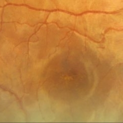
A 17-year-old healthy woman noticed a pacman-shaped scotoma in her temporal right vision. Acuity measured 20/20 and color vision measured 11/11. High magnification of the color fundus photograph showing some possible pigmentary changes at the fovea.
Photographer: Geoffrey Emerson, MD, PhD, Retina Center, Minneapolis
Condition/keywords: scotoma
-
---thumb.jpg/image-square;max$300,300.ImageHandler) Terson's Syndrome
Terson's Syndrome
Oct 8 2013 by Maurice F. Rabb
39 year female with a long history of chronic back pain treated by a sequence of epidural injections. Following her last injection, she complained of a moderately severe protracted headache and had several attempts at placement of an epidural blood patch without success. Under general anesthesia, she underwent injection of a larger volume of saline in an attempt to stem a presumed CSF leak producing "spinal headache". In the left eye she demonstrated multiple superficial and deep intraretinal hemorrhages associated with mild disc swelling and a central scotoma. In the right eye she showed a posterior subhyaloid and sub-internal limiting membrane hemorrhage with buffy coat layering superiorly. The visual acuity measured hand motions OD, 20/200 OS. The patient underwent a surgical evacuation of the sub-ILM hemorrhage.
Condition/keywords: Terson's Syndrome
-
 Retinal Pigment Changes After Blunt Ocular Trauma
Jun 27 2016 by Rita Couceiro, MD, MS
A 19-year-old man suffered blunt trauma of the left eye with a ball during soccer practice. At day 3 after trauma (upper pictures) the retinal area superior to the fovea looked pale and visual acuity was reduced to 20/32. This area revealed hypersignaling of retinal layers on OCT and the foveal area showed a localized disruption of retinal layers above the RPE. At day 30 (lower pictures) the retinal area of pallor showed pigmentary changes and OCT revealed atrophy of the external retinal layers. However the localized subfoveal retinal disruption was improved and only a slight disruption was seen on OCT at the ellipsoid level. Visual acuity of the left eye was restored to 20/20 although a scotoma remained.
Photographer: Rita Couceiro, Serviço de Oftalmologia do Hospital de Santa Maria, Lisboa, Portugal
Condition/keywords: blunt trauma, commotio retinae, pigment changes
-
 AZOOR
AZOOR
Aug 24 2012 by Geoffrey G. Emerson, MD, PhD, FASRS
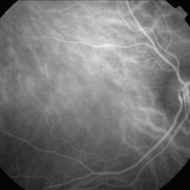
A 17-year-old healthy woman noticed a pacman-shaped scotoma in her temporal right vision. Acuity measured 20/20 and color vision measured 11/11. Indocyanine angiography was unremarkable.
Photographer: Geoffrey Emerson, MD, PhD, Retina Center, Minneapolis
Condition/keywords: scotoma
-
---thumb.jpg/image-square;max$300,300.ImageHandler) Cilioretinal Artery Occlusion with Central Retinal Vein Occlusion
Cilioretinal Artery Occlusion with Central Retinal Vein Occlusion
Mar 9 2013 by Gabriela Lopezcarasa Hernandez, MD
A 46-year-old male with decrease in visual acuity in left eye and central scotoma.
Photographer: Araceli Rojas Arriaga, Hospital Angeles Lomas, Mexico
Imaging device: Zeiss FF4
Condition/keywords: central retinal vein occlusion (CRVO), cilioretinal artery occlusion
-
---thumb.jpg/image-square;max$300,300.ImageHandler) Subretinal Hemorrhage Secondary to Macroaneurysm
Subretinal Hemorrhage Secondary to Macroaneurysm
Jul 24 2013 by Gabriela Lopezcarasa Hernandez, MD
65-year-old female patient with sudden scotoma in right eye who was previously diagnosed with macroaneurysm in temporal superior arcade.
Photographer: Araceli Rojas Arriaga, Hospital Angeles Lomas, Mexico
Imaging device: ZEISS FF4
Condition/keywords: macroaneurysm, subretinal hemorrhage
-
 AZOOR
AZOOR
Aug 24 2012 by Geoffrey G. Emerson, MD, PhD, FASRS
A 17-year-old healthy woman noticed a pacman-shaped scotoma in her temporal right vision. Acuity measured 20/20 and color vision measured 11/11. There was an enlarged physiologic blind spot on Humphry visual field testing. The fellow eye was normal.
Photographer: Geoffrey Emerson, MD, PhD, Retina Center, Minneapolis
Condition/keywords: scotoma
-
---thumb.jpg/image-square;max$300,300.ImageHandler) Subretinal Hemorrhage Secondary to Macroaneurysm
Subretinal Hemorrhage Secondary to Macroaneurysm
Jul 24 2013 by Gabriela Lopezcarasa Hernandez, MD
65-year-old female patient with sudden scotoma in right eye who was previously diagnosed with macro aneurysm in temporal superior arcade.
Photographer: Araceli Rojas Arriaga, Hospital Angeles Lomas, Mexico
Imaging device: ZEISS FF4
Condition/keywords: macroaneurysm, subretinal hemorrhage
-
---thumb.jpg/image-square;max$300,300.ImageHandler) Central Scotoma
Central Scotoma
 Loading…