Search results (171 results)
-
 Acute Posterior Multifocal Placoid Pigment Epitheliopathy
Sep 15 2012 by Roy D. Brod, MD
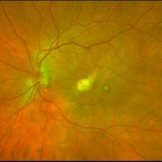
Fundus photograph left eye demonstrating cream colored placoid lesions in 28-year-old male patient with 4-day history of multiple scotomas OU.
Photographer: Julia Walker
Condition/keywords: acute posterior multifocal placoid pigment epitheliopathy (APMPPE), placoid retinal lesions, scotoma
-
 Acute Posterior Multifocal Placoid Pigment Epitheliopathy
Sep 15 2012 by Roy D. Brod, MD
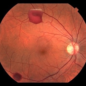
Fundus photograph right eye demonstrating cream colored placoid lesions in 28-year-old male patient with 4-day history of multiple scotomas OU.
Photographer: Julia Walker
Condition/keywords: acute posterior multifocal placoid pigment epitheliopathy (APMPPE), placoid retinal lesions, scotoma
-
 AZOOR
AZOOR
Aug 24 2012 by Geoffrey G. Emerson, MD, PhD, FASRS
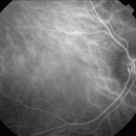
A 17-year-old healthy woman noticed a pacman-shaped scotoma in her temporal right vision. Acuity measured 20/20 and color vision measured 11/11. Indocyanine angiography was unremarkable.
Photographer: Geoffrey Emerson, MD, PhD, Retina Center, Minneapolis
Condition/keywords: scotoma
-
 AZOOR
AZOOR
Aug 24 2012 by Geoffrey G. Emerson, MD, PhD, FASRS
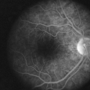
A 17-year-old healthy woman noticed a pacman-shaped scotoma in her temporal right vision. Acuity measured 20/20 and color vision measured 11/11. Angiography showed some late staining of the nasal macula.
Photographer: Geoffrey Emerson, MD, PhD, Retina Center, Minneapolis
Condition/keywords: scotoma
-
 AZOOR
AZOOR
Aug 24 2012 by Geoffrey G. Emerson, MD, PhD, FASRS
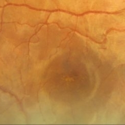
A 17-year-old healthy woman noticed a pacman-shaped scotoma in her temporal right vision. Acuity measured 20/20 and color vision measured 11/11. High magnification of the color fundus photograph showing some possible pigmentary changes at the fovea.
Photographer: Geoffrey Emerson, MD, PhD, Retina Center, Minneapolis
Condition/keywords: scotoma
-
 AZOOR
AZOOR
Aug 24 2012 by Geoffrey G. Emerson, MD, PhD, FASRS
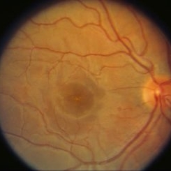
A 17-year-old healthy woman noticed a pacman-shaped scotoma in her temporal right vision. Acuity measured 20/20 and color vision measured 11/11. Possible pigmentary changes were visible at the fovea.
Photographer: Geoffrey Emerson, MD, PhD, Retina Center, Minneapolis
Condition/keywords: scotoma
-
 AZOOR
AZOOR
Aug 24 2012 by Geoffrey G. Emerson, MD, PhD, FASRS
A 17-year-old healthy woman noticed a pacman-shaped scotoma in her temporal right vision. Acuity measured 20/20 and color vision measured 11/11. Multifocal ERG showed decreased signal in the temporal right vision
Photographer: Geoffrey Emerson, MD, PhD, Retina Center, Minneapolis
Condition/keywords: scotoma
-
 AZOOR
AZOOR
Aug 24 2012 by Geoffrey G. Emerson, MD, PhD, FASRS
A 17-year-old healthy woman noticed a pacman-shaped scotoma in her temporal right vision. Acuity measured 20/20 and color vision measured 11/11. There was an enlarged physiologic blind spot on Humphry visual field testing. The fellow eye was normal.
Photographer: Geoffrey Emerson, MD, PhD, Retina Center, Minneapolis
Condition/keywords: scotoma
-
 Endogenous Endophthalmitis With Suspected Systemic Candidiasis
Jan 9 2018 by Olivia Rainey
Ultra-wide field Optos pseudocolor image of an 45-year-old male presenting with endogenous endophthalmitis affecting his left eye. Candidiasis was at high considerations due to intravenous drug abuse and recent history of dental abscess. Patient developed a subretinal infiltrate resulting in central scotoma and responded well to anti-fungal treatment.
Photographer: Olivia Rainey
Imaging device: Optos California
Condition/keywords: candida endophthalmitis, drug abuse, endogenous endophthalmitis, left eye, Optos, pseudocolor, retinal infiltrates, scotoma, ultra-wide field imaging
-
 Susac's syndrome
Dec 5 2017 by Marina Gilca, MD
Fluorescein angiogram of a 33 year-old female presenting with recurrent scotomas OS.
Photographer: Retina Consultants LTD
Condition/keywords: scotoma
-
 Syphilitic Optic Neuritis Acute Humphrey VF Scotomas
Oct 11 2012 by Jeffrey G. Gross, MD, FASRS
Syphilitic optic neuritis, acute Humphry VF, scotomas.
Condition/keywords: acute humphry VF, scotoma, syphilitic optic neuritis
-
 A Motor Vehicle Accident Causing Valsalva Retinopathy OD, While Racing A Side By Side 4 Wheel Off-Road Vehicle
Apr 29 2020 by John S. King, MD
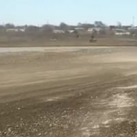
43-year-old white male who was injured while racing a side by side 4-wheel off-road vehicle (see Video: https://imagebank.asrs.org/file/53854/sxs-crash-during-a-race-causing-valsalva-retinopathy-od). He presented about three weeks after the injury. He was being seen by his local eye doctor who wanted an evaluation for the retinal heme and scotoma. His main complaint was a central/parcentral scotoma described as a greyish area in vision. Va 20/50 OD, nomotensive, no APD (by technician), anterior segment u/r; see picture for the fundus exam - of note there are superficial/preretinal heme, with layering of the heme superiorly, and small superficial heme at nasal edge of the optic disc; in the parafoveal region nasally there is some mottling of the RPE that may indicate an area of prior commotio retinae (also possible to have TON), which may account for his scotoma. Really bad accident (video), and amazingly, he had no LOC or injuries other than the right retina. Helmet and racing harness seat belt were used.
Photographer: Asli Ahmed
Imaging device: Topcon 50
Condition/keywords: valsalva retinopathy
-
 A Motor Vehicle Accident Causing Valsalva Retinopathy OD, While Racing A Side By Side 4 Wheel Off-Road Vehicle
May 5 2020 by John S. King, MD
A 43-year-old white male who was injured while racing his side by side 4 wheel off-road vehicle (this is a video he showed me on his phone). He presented about three weeks after the injury. He was being seen by his local eye doctor who wanted an evaluation for the retinal heme and scotoma. His main complaint was a central/parcentral scotoma described as a greyish area in vision. Va 20/50 OD, nomotensive, no APD (by technician), anterior segment u/r; see {https://imagebank.asrs.org/file/53828/sxs-crash-during-a-race-causing-valsalva-retinopathy-od} for the fundus exam - of note there are superficial/preretinal heme, with layering of the heme superiorly; in the parafoveal region nasally there is some mottling of the RPE that may indicate an area of prior commotio retinae (also possible to have TON), which may account for his scotoma. Really bad accident, and amazingly, he had no LOC or injuries other than the right retina. Helmet and racing harness seat belt were used.
Condition/keywords: motor vehicle accident, trauma, valsalva retinopathy
-
 Acute Compressive Optic Neuropathy
Jun 1 2019 by John S. King, MD
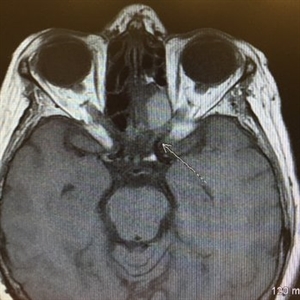
84-year-old white female with acute loss of vision in the left eye one day ago was sent here after going to the ED per primary eye provider. She described vision loss as a grey curtain that became total darkness. She had left sided temporal tenderness and some left sided neck pain. In the ED the cardiac work-up was u/r, the ESR and CRP were normal, and the CTH showed some non-specific opacification in the L ethmoid sinus. Acuity was HM OS with RAPD, normal EOMs, no proptosis or ptosis, posteriorly no SVPs were noted; the optic discs were pink and flat; no emboli or retinal whitening present; some bear tracks located nasally (see photo). She was referred to Dr. Doyle, who ordered an MRI, which showed a large mucocele with bony erosion into the left orbit, along with some ON enhancement possibly from compression (see images). She was operated that night and later recovered to 20/40 in that eye with a residual, inferior arcuate scotoma.
Condition/keywords: bear tracks, optic neuropathy
-
 Acute Macular Neuroretinopathy
Apr 12 2021 by Iuri Golubev, MD
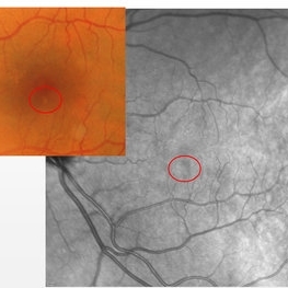
46-year-old female with sudden onset paracentral scotoma below the central point of fixation in her left eye. Enface image shows a wedge shaped lesion pointing towards the fovea (top left). The lesion was spanning outer retinal layers from OPL to RPE (top left insert). One month later, the lesion has diminished in size, and was only involving retinal layers from ellipsoid zone to RPE(top right). At 4 months since presentation, the patient did not have any signs of AMN identifiable on enface or b-scan images (bottom center). Patient's symptoms has slowly improved and eventually resolved over the course of the next 4 years.
Imaging device: Zeiss Cirrus 5000
Condition/keywords: acute macular neuroretinopathy, acute macular outer retinopathy
-
 Acute Macular Neuroretinopathy OD
Mar 28 2013 by John S. King, MD
19-year-old patient c ADHD and acute photopsia following flu-like illness; inferior scotoma OD. Ellipsoid region abn c characteristic parafoveal lesions on voxel.
Photographer: Wayne A. Ladlee Jr., OcuSight Eye Care Center, Rochester, NY
Imaging device: Cirrus
Condition/keywords: acute macular neuroretinopathy
-
 Acute Macular Neuroretinopathy OS
Mar 28 2013 by John S. King, MD
19-year-old patient c ADHD and acute photopsia following flu-like illness; inferior scotoma OD. Ellipsoid region abn c characteristic parafoveal lesions on voxel.
Photographer: Wayne A. Ladlee Jr., OcuSight Eye Care Center, Rochester, NY
Imaging device: Cirrus
Condition/keywords: acute macular neuroretinopathy
-
 Acute Optic Neuropathy Due to Large Mucocele
Jun 1 2019 by John S. King, MD
84-year-old white female with acute loss of vision in the left eye one day ago was sent here after going to the ED per primary eye provider. She described vision loss as a grey curtain that became total darkness. She had left sided temporal tenderness and some left sided neck pain. In the ED the cardiac work-up was u/r, the ESR and CRP were normal, and the CTH showed some non-specific opacification in the L ethmoid sinus. Acuity was HM OS with RAPD, normal EOMs, no proptosis or ptosis, posteriorly no SVPs were noted; the optic discs were pink and flat; no emboli or retinal whitening present; some bear tracks located nasally (see photo). She was referred to Dr. Doyle, who ordered an MRI, which showed a large mucocele with bony erosion into the left orbit, along with some ON enhancement possibly from compression (see Images). She was operated that night and later recovered to 20/40 in that eye with a residual, inferior arcuate scotoma.
Condition/keywords: bear tracks, optic neuropathy
-
 Acute Optic Neuropathy Due to Large Mucocele (Incidental Bear Tracks)
Jun 1 2019 by John S. King, MD
84-year-old white female with acute loss of vision in the left eye one day ago was sent here after going to the ED per primary eye provider. She described vision loss as a grey curtain that became total darkness. She had left sided temporal tenderness and some left sided neck pain. In the ED the cardiac work-up was u/r, the ESR and CRP were normal, and the CTH showed some non-specific opacification in the L ethmoid sinus. Acuity was HM OS with RAPD, normal EOMs, no proptosis or ptosis, posteriorly no SVPs were noted; the optic discs were pink and flat; no emboli or retinal whitening present; some bear tracks located nasally (see photo). She was referred to Dr. Doyle, who ordered an MRI, which showed a large mucocele with bony erosion into the left orbit, along with some ON enhancement possibly from compression (see images). She was operated that night and later recovered to 20/40 in that eye with a residual, inferior arcuate scotoma.
Photographer: Karin Aletter
Imaging device: Topcon 50
Condition/keywords: bear tracks, optic neuropathy
-
 Acute syphilitic posterior placoid chorioretinitis
Apr 24 2022 by Aniruddha K Agarwal, MD
Green-light fundus autofluorescence (FAF) of the right eye from a 55-year-old man with risk factors for sexually trasnmitted diseases who presented to the retina clinic for a central scotoma. Funduscopy revealed a placoid lesion in the posterior pole. FAF highlights a hyperautofluorescent placoid lesion involving the macula with granular hyperfluorescence. The patient tested positive for syphilis and received intravenous penicillin treatment.
Photographer: Esther CIANCAS, MD, PhD, Gema CRESPO-RODRÍGUEZ, RN
Imaging device: Zeiss Clarus fundus camera
Condition/keywords: chorioretinitis, IUSG, syphilis, uveitis
-
 AMN (Acute Macular Neuroretinitis) #2
Apr 28 2019 by Niloofar Piri, MD
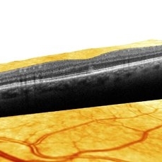
HD-OCT image of 53-year-old man who presented with a superior small paracentral scotoma for 1 month. He had very small hypopigmented area inferior to the fovea and hyporeflectivity on NIR image ( #1). OCT demonstrated a vertical hyper-reflective band extending from OPL to RPE. This form is type 2 AMN which is due to occlusion of deep capillary plexus.
Photographer: Niloofar Piri,MD
Condition/keywords: acute macular neuroretinitis, acute macular neuroretinopathy
-
 AMN (Acute Macular Neurortinitis)
Apr 28 2019 by Niloofar Piri, MD
HD-OCT image of 53-year-old man who presented with a superior small paracentral scotoma for 1 month. He had very small hypopigmented area inferior to the fovea and hyporeflectivity on NIR image ( #1). OCT demonstrated a vertical hyper-reflective band extending from OPL to RPE. This form is type 2 AMN which is due to occlusion of deep capillary plexus. #2
Condition/keywords: acute macular neuroretinitis, acute macular neuroretinopathy
-
 Best Disease
Apr 24 2024 by Marcelo Zas, MD PhD
Best vitelliform macular dystrophy (BVMD) or Best disease. Is the most common autosomal dominant macular dystrophy. It involves the retinal pigment epithelium (RPE), and leads to a characteristic bilateral yellow “egg-yolk” appearance of the macula as you can see in this image. Essentially, BVMD is considered to have 6 clinical stages: Previtelliform, Vitelliform, Pseudohypopyon, Vitelleruptive, Atrophic and Choroidal neovascularization. As the disease progresses, patients may experience a slow, bilateral decrease in visual acuity, central scotoma, or metamorphopsia. With secondary CNV, visual decline can be rapid, however.
Photographer: Luciano Scorsetti MD
Condition/keywords: Macular Dystrophy
-
 Branch Retinal Artery Occlusion
Sep 9 2018 by Gabriela Lopezcarasa Hernandez, MD
88-year-old female patient with sudden decrease in visual acuity and scotoma in left eye, please notice the widening retina due to retinal edema of branch occlusion with hollenhorst plaque in the artery and the optic nerve.
Photographer: Araceli Rojas
Imaging device: Zeiss FF4
Condition/keywords: branch retinal artery occlusion (BRAO)
-
 Branch Retinal Artery Occlusion
Sep 9 2018 by Gabriela Lopezcarasa Hernandez, MD
88-year-old female patient with sudden decrease in visual acuity and scotoma in left eye, please notice the branch occlusion with hollenhorst plaque and the delay perfusion in the involved arteria.
Photographer: Araceli Rojas
Imaging device: Zeiss FF4
Condition/keywords: branch retinal artery occlusion (BRAO)
 Loading…