 Initializing download.
-
 By Rui Zhang, BA
Dartmouth, Geisel School of Medicine
Co-author(s): Dimosthenis Mantopoulos, MD PhD (Dartmouth-Hitchcock Medical Center) - Uploaded on Apr 15, 2021.
- Last modified by Caroline Bozell on Apr 15, 2021.
- Rating
- Appears in
- Choroidal MRSA Abscess
- Condition/keywords
- abscess, MRSA sepsis, acute leukemia
- Photographer
- Raymond Mok, CRA COMT (Dartmouth-Hitchcock Medical Center)
- Imaging device
-
Fundus camera
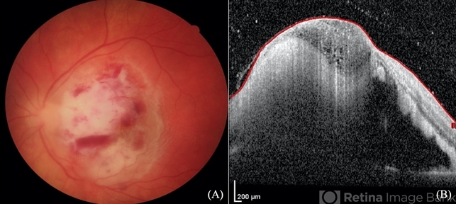
Optical coherence tomography - Description
- A 14-year-old boy receiving induction chemotherapy for acute lymphocytic leukemia (ALL) complained of floaters and central scotoma in his left eye. (A) Fundus photography showed sub-macular choroidal abscess with intraretinal hemorrhage and edema. (B) OCT confirmed that the abscess had not penetrated the retinal pigment epithelium (RPE). Due to systemic septic signs (fever, tachycardia, tachypnea, new-onset papules), blood cultures were drawn and they came back positive for methicillin-resistant staphylococcus aureus (MRSA). Patient was promptly treated with both IV and intravitreal antibiotics. This is a case of sub-macular choroidal MRSA abscess in the setting of systemic bacteremia in an immunocompromised host.


