 Initializing download.
-
 By Iuri Golubev, MD
- Uploaded on Apr 12, 2021.
- Last modified by Caroline Bozell on Apr 15, 2021.
- Rating
- Appears in
- AZOOR
- Condition/keywords
- acute macular neuroretinopathy, acute macular outer retinopathy
- Imaging device
-
Optical coherence tomography system
Zeiss Cirrus 5000 - Description
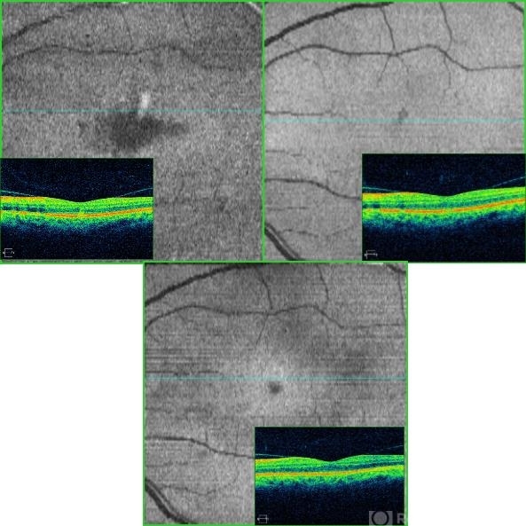
- 46-year-old female with sudden onset paracentral scotoma below the central point of fixation in her left eye. Enface image shows a wedge shaped lesion pointing towards the fovea (top left). The lesion was spanning outer retinal layers from OPL to RPE (top left insert). One month later, the lesion has diminished in size, and was only involving retinal layers from ellipsoid zone to RPE(top right). At 4 months since presentation, the patient did not have any signs of AMN identifiable on enface or b-scan images (bottom center). Patient's symptoms has slowly improved and eventually resolved over the course of the next 4 years.


---thumb.jpg/image-square;max$79,0.ImageHandler "AMN")
---thumb.jpg/image-square;max$79,0.ImageHandler "AMN, Red Free")
---thumb.jpg/image-square;max$79,0.ImageHandler "AMN, FA (OS)")
---thumb.jpg/image-square;max$79,0.ImageHandler "AMN, FA (OS)")
---thumb.jpg/image-square;max$79,0.ImageHandler "AMN Acute Macular Neuroretinopathy")