Search results (171 results)
-
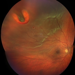
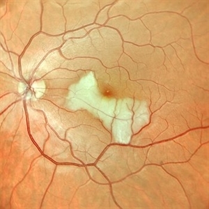
 MEWDS
MEWDS
Oct 9 2020 by David L Kilpatrick, MD
26-year-old female presented with unilateral vision loss. She c/o flashes and a peripheral scotoma. Vision was 20/100. On exam, she showed foveal granularity, mild disc edema, and white dots as seen. Three weeks later, white dots had resolved and vision improved to 20/25.
Photographer: Mississippi Retina Associates
Imaging device: Optos
Condition/keywords: multiple evanescent white dot syndrome (MEWDS)
-
 Rhegmatogenous Retinal Detachment
Mar 3 2021 by Patrik Rajs
A 51-year-old female patient presented with inferior nasal scotoma and 5/10 vision in the right eye due to a retinal detachment with a giant retinal horseshoe tear.
Photographer: Patrik Rajs, EYE CLINIC of Jan Evangelista Purkyne University and Masaryk Hospital, Czech Republic, Ústí nad Labem
Imaging device: Clarus 700
Condition/keywords: giant retinal tear
-
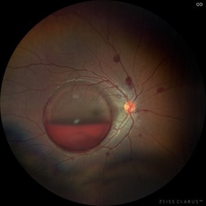
 Acute syphilitic posterior placoid chorioretinitis
Apr 24 2022 by Aniruddha K Agarwal, MD
Green-light fundus autofluorescence (FAF) of the right eye from a 55-year-old man with risk factors for sexually trasnmitted diseases who presented to the retina clinic for a central scotoma. Funduscopy revealed a placoid lesion in the posterior pole. FAF highlights a hyperautofluorescent placoid lesion involving the macula with granular hyperfluorescence. The patient tested positive for syphilis and received intravenous penicillin treatment.
Photographer: Esther CIANCAS, MD, PhD, Gema CRESPO-RODRÍGUEZ, RN
Imaging device: Zeiss Clarus fundus camera
Condition/keywords: chorioretinitis, IUSG, syphilis, uveitis
-
 AMN (Acute Macular Neuroretinitis) #2
Apr 28 2019 by Niloofar Piri, MD
HD-OCT image of 53-year-old man who presented with a superior small paracentral scotoma for 1 month. He had very small hypopigmented area inferior to the fovea and hyporeflectivity on NIR image ( #1). OCT demonstrated a vertical hyper-reflective band extending from OPL to RPE. This form is type 2 AMN which is due to occlusion of deep capillary plexus.
Photographer: Niloofar Piri,MD
Condition/keywords: acute macular neuroretinitis, acute macular neuroretinopathy
-
 AMN (Acute Macular Neurortinitis)
Apr 28 2019 by Niloofar Piri, MD
HD-OCT image of 53-year-old man who presented with a superior small paracentral scotoma for 1 month. He had very small hypopigmented area inferior to the fovea and hyporeflectivity on NIR image ( #1). OCT demonstrated a vertical hyper-reflective band extending from OPL to RPE. This form is type 2 AMN which is due to occlusion of deep capillary plexus. #2
Condition/keywords: acute macular neuroretinitis, acute macular neuroretinopathy
-
 Branch Retinal Artery Occlusion
Sep 9 2018 by Gabriela Lopezcarasa Hernandez, MD
88-year-old female patient with sudden decrease in visual acuity and scotoma in left eye, please notice the widening retina due to retinal edema of branch occlusion with hollenhorst plaque in the artery and the optic nerve.
Photographer: Araceli Rojas
Imaging device: Zeiss FF4
Condition/keywords: branch retinal artery occlusion (BRAO)
-
 Branch Retinal Artery Occlusion
Sep 9 2018 by Gabriela Lopezcarasa Hernandez, MD
88-year-old female patient with sudden decrease in visual acuity and scotoma in left eye, please notice the branch occlusion with hollenhorst plaque and the delay perfusion in the involved arteria.
Photographer: Araceli Rojas
Imaging device: Zeiss FF4
Condition/keywords: branch retinal artery occlusion (BRAO)
-
 Central Retinal Vein Occlusion
Feb 25 2025 by Prithvi Chandrakanth
A 61-year-old woman with a history of hypertension noticed a sudden painless blurring of vision in her left eye. Over the next few days, the blurriness persisted, and she experienced a mild central scotoma. On examination, Fundoscopic evaluation revealed dilated, tortuous retinal veins, retinal hemorrhages, and macular oedema.
Photographer: DR.PRITHVI CHANDRAKANTH, DR.CHANDRAKANTH NETHRALAYA, KOZHIKODE
Imaging device: EIDON
Condition/keywords: CRVO, CRVO WITH MACULA EDEMA, flame shaped retinal hemorrhage
-
 Cilioretinal Artery Occlusion
May 14 2024 by Eloy Mata-Cortes, MD
Color image capturing the left eye of a 32-year-old female. Despite a negative ophthalmological and medical history, she reported three days of blurred vision and a paracentral scotoma in her left eye, while maintaining central vision. The image reveals retinal whitening, extends from the parafoveal region to the inferotemporal arcade indicative of cilioretinal artery occlusion. Following this observation, the patient was referred for systemic assessment to explore the underlying etiology of the occlusion.
Photographer: Eloy Mata-Cortes, MD, Instituto Mexicano de Oftalmología, Querétaro, México
Imaging device: Nidek Mirante
Condition/keywords: cilioretinal artery occlusion, oclussion, retinal whitening
-
---thumb.jpg/image-square;max$300,300.ImageHandler) Cilioretinal Artery Occlusion with Central Retinal Vein Occlusion
Cilioretinal Artery Occlusion with Central Retinal Vein Occlusion
Mar 9 2013 by Gabriela Lopezcarasa Hernandez, MD
A 46-year-old male with decrease in visual acuity in left eye and central scotoma.
Photographer: Araceli Rojas Arriaga, Hospital Angeles Lomas, Mexico
Imaging device: Zeiss FF4
Condition/keywords: central retinal vein occlusion (CRVO), cilioretinal artery occlusion
-
---thumb.jpg/image-square;max$300,300.ImageHandler) Cilioretinal Artery Occlusion with Central Retinal Vein Occlusion
Cilioretinal Artery Occlusion with Central Retinal Vein Occlusion
Mar 9 2013 by Gabriela Lopezcarasa Hernandez, MD
A 46-year-old male with decrease in visual acuity in left eye and central scotoma.
Photographer: Araceli Rojas Arriaga, Hospital Angeles Lomas, Mexico
Imaging device: Zeiss FF4
Condition/keywords: central retinal artery occlusion (CRAO), central retinal vein occlusion (CRVO), cilioretinal artery occlusion
-
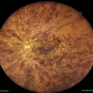
 Myelinated Nerve Fibers
Apr 18 2025 by DR Rohit Gupta
The **myelinated nerve fibers of the optic disc** (also known as **medullated nerve fibers**) are retinal nerve fibers that retain their myelin sheath as they pass through the optic nerve head. Normally, retinal nerve fibers are unmyelinated to allow for light transparency, but in some cases, myelination extends anteriorly into the retina, appearing as a striking white, feathery patch on the optic disc or peripapillary retina. ### **Key Features:** 1. **Appearance:** - Dense, white, striated patches with feathery edges. - Typically located at the superior or inferior pole of the optic disc. - May obscure retinal vessels underneath. 2. **Clinical Significance:** - Usually **benign** and asymptomatic. - **Congenital** (present at birth or early childhood). - Rarely associated with **visual field defects** (e.g., scotomas corresponding to the area of myelination). - Occasionally linked with **high myopia** or **amblyopia** if extensive. 3. **Pathophysiology:** - Failure of oligodendrocytes or Schwann cells to stop myelination at the lamina cribrosa. - Normally, myelination stops at the optic nerve head, but in this condition, it extends into the retina. 4. **Diagnosis:** - **Fundoscopy:** Classic white, feathery appearance. - **Optical Coherence Tomography (OCT):** Shows thickened retinal nerve fiber layer (RNFL). - **Visual Field Testing:** May detect defects if large. 5. **Differential Diagnosis:** - Optic disc edema - Cotton wool spots - Retinoblastoma (rarely, but must be ruled out in children) 6. **Management:** - No treatment required if asymptomatic. - Monitor for amblyopia in children. - Rare cases with significant visual impairment may need further evaluation. ### **Fun Fact:** Myelinated nerve fibers are seen in **~0.5-1%** of the population and are usually an incidental finding.
Photographer: Dr Rohit gupta
Imaging device: Samsung S21
Condition/keywords: Medulated Nerve fibre, Medullated Nerve fibres, myelinated nerve fibers, Myelinated Nerve Fibres, optic disc drusen
-
 Preeclampsia in a 30-Year-Old - Red Free Photograph - RE
Nov 25 2015 by Roy Schwartz, MD
A 30-year-old presented with central scotoma and blurred vision a day following C-section for preeclampsia.
Photographer: Galit Yair Pur
Condition/keywords: blurred vision, central scotoma, preeclampsia
-
 RAMA with Sub ILM Hemorrhage
Jan 31 2018 by John S. King, MD
73-year-old with well controlled diabetes and hypertension presented with a month onset of acute central scotoma; CF 5'
Photographer: Stacey
Imaging device: Cirrus
Condition/keywords: ruptured macroaneurysm, sub-inner limiting membrane hemorrhage
-
 Susac's Syndrome
Feb 13 2018 by John S. King, MD
Background: 46-year-old WF with CML (stable on Sprycel) saw her PCP for headaches without known cause; Headaches worsened and became confused, disoriented, off balance, and impaired short term memory. Heme-oncology ordered MRI that showed abnormal signal in the cerebellum and other parts of the brain, and LP has elevated protein. LP did show positive tau test, but fortunately, was a false positive for CJD. IV and PO steroids started and symptoms improved. MRI showed much improvement one month since starting steroids. 3 weeks later had a scotoma in right eye and eye doctor did not find anything at that time to cause it. Tinnitus developed (and some intermittent vertigo before that) and ENT referred back to eye doctor, who then referred the patient to Dr. Zocchi. He found a CWS and BRAO OD, and bilateral arteritis. She had some additional work-up for vasculitis. Given the retinal arteritis, cochlear issues, and MRI findings, Dr.Zocchi suspected Susac's Syndrome. She was started on multiple regimens including prednisone, IVIG, azathiprine, and MTX, and has had the best reponse to IVIG (FA shows a recurrence/worsening while adjusting IMT). She is stable and doing well with 20/20 vision in both eyes.
Photographer: Kay Dalby
Imaging device: Topcon
Condition/keywords: retinal vasculitis, Susac's syndrome
-
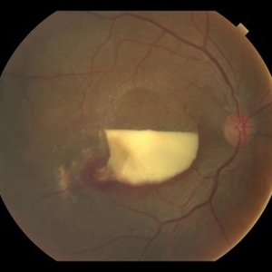
 Valsalva Retinopathy
Nov 18 2022 by Niloofar Piri, MD
21 yo female presented with decaresed central vision and scotoma immediately after severe vomiting. Color fundus phtograph demonstrates large sub ILM layered hemorrhage in the macula consistent with valsalva retinopathy. Notice the sacttered blot retinal hemorrhages in mid-periphery.
Photographer: Rocio Bentivegna, MD, Saint Louis University
Condition/keywords: sub ILM hemorrhage, valsalva retinopathy
-
 Rhegmatogenous Retinal Detachment
Aug 23 2012 by Gabriela Lopezcarasa Hernandez, MD
30-year-old male with floaters and inferonasal scotoma.
Photographer: Gabriela Lopezcarasa Hernandez, Hospital Angeles Lomas
Imaging device: ZEISS FF4
Condition/keywords: floaters, inferonasal scotoma
-
 A Motor Vehicle Accident Causing Valsalva Retinopathy OD, While Racing A Side By Side 4 Wheel Off-Road Vehicle
Apr 29 2020 by John S. King, MD
43-year-old white male who was injured while racing a side by side 4-wheel off-road vehicle (see Video: https://imagebank.asrs.org/file/53854/sxs-crash-during-a-race-causing-valsalva-retinopathy-od). He presented about three weeks after the injury. He was being seen by his local eye doctor who wanted an evaluation for the retinal heme and scotoma. His main complaint was a central/parcentral scotoma described as a greyish area in vision. Va 20/50 OD, nomotensive, no APD (by technician), anterior segment u/r; see picture for the fundus exam - of note there are superficial/preretinal heme, with layering of the heme superiorly, and small superficial heme at nasal edge of the optic disc; in the parafoveal region nasally there is some mottling of the RPE that may indicate an area of prior commotio retinae (also possible to have TON), which may account for his scotoma. Really bad accident (video), and amazingly, he had no LOC or injuries other than the right retina. Helmet and racing harness seat belt were used.
Photographer: Asli Ahmed
Imaging device: Topcon 50
Condition/keywords: valsalva retinopathy
-
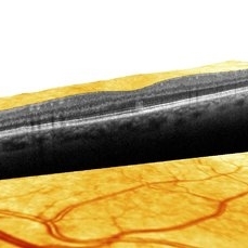
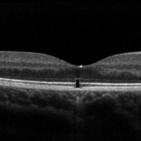 Laser Pointer Maculopathy
Apr 28 2019 by Bastián Schmidt Arias
Optical coherence tomography of a 13-year-old adolescent with a laser pointer maculopathy, 20/20 visual acuity with paracentral scotoma.
Photographer: Bastian Schmidt
Imaging device: Topcon 3D OCT-2000
Condition/keywords: laser pointer maculopathy, maculopathy, optical coherence tomography (OCT)
-
 A Motor Vehicle Accident Causing Valsalva Retinopathy OD, While Racing A Side By Side 4 Wheel Off-Road Vehicle
May 5 2020 by John S. King, MD
A 43-year-old white male who was injured while racing his side by side 4 wheel off-road vehicle (this is a video he showed me on his phone). He presented about three weeks after the injury. He was being seen by his local eye doctor who wanted an evaluation for the retinal heme and scotoma. His main complaint was a central/parcentral scotoma described as a greyish area in vision. Va 20/50 OD, nomotensive, no APD (by technician), anterior segment u/r; see {https://imagebank.asrs.org/file/53828/sxs-crash-during-a-race-causing-valsalva-retinopathy-od} for the fundus exam - of note there are superficial/preretinal heme, with layering of the heme superiorly; in the parafoveal region nasally there is some mottling of the RPE that may indicate an area of prior commotio retinae (also possible to have TON), which may account for his scotoma. Really bad accident, and amazingly, he had no LOC or injuries other than the right retina. Helmet and racing harness seat belt were used.
Condition/keywords: motor vehicle accident, trauma, valsalva retinopathy
-
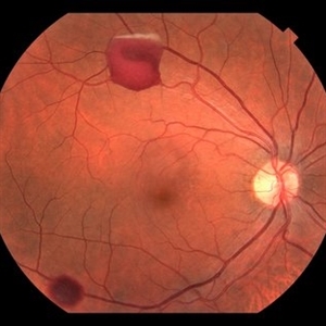
 Acute Posterior Multifocal Placoid Pigment Epitheliopathy
Sep 15 2012 by Roy D. Brod, MD
Fundus photograph left eye demonstrating cream colored placoid lesions in 28-year-old male patient with 4-day history of multiple scotomas OU.
Photographer: Julia Walker
Condition/keywords: acute posterior multifocal placoid pigment epitheliopathy (APMPPE), placoid retinal lesions, scotoma
-
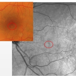
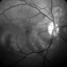
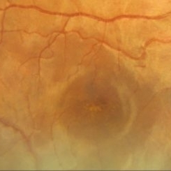 AZOOR
AZOOR
Aug 24 2012 by Geoffrey G. Emerson, MD, PhD, FASRS
A 17-year-old healthy woman noticed a pacman-shaped scotoma in her temporal right vision. Acuity measured 20/20 and color vision measured 11/11. High magnification of the color fundus photograph showing some possible pigmentary changes at the fovea.
Photographer: Geoffrey Emerson, MD, PhD, Retina Center, Minneapolis
Condition/keywords: scotoma
-
 Cilioretinal Artery Occlusion with Central Retinal Vein Occlusion
Mar 9 2013 by Gabriela Lopezcarasa Hernandez, MD
A 46-year-old male with decrease in visual acuity in left eye and central scotoma.
Photographer: Araceli Rojas Arriaga, Hospital Angeles Lomas, Mexico
Imaging device: ZEISS FF4
Condition/keywords: cilioretinal artery occlusion
-
 MEWDS
MEWDS
Feb 14 2018 by Daniel K Bennett, MD
24-year-old white female with mild scotoma.
Imaging device: Zeiss Visucam
Condition/keywords: multiple evanescent white dot syndrome (MEWDS)
-
 Plaquenil Toxicity
Apr 30 2013 by Theodore Leng, MD, MS, FASRS
Right eye 10-2 HVF from a 44-year-old woman with bilateral plaquenil toxicity. A ring scotoma is present.
Condition/keywords: hydroxychloroquine toxicity, plaquenil toxicity
 Loading…