 Initializing download.
-
 By Niloofar Piri, MD
SSM Health Group, St Louis University
Co-author(s): Jillian baker, DO, Saint Louis University; Sean Kelso, Saint Louis University - Uploaded on Mar 31, 2023.
- Last modified by Joshua Friedman on Apr 3, 2023.
- Rating
- Appears in
- Miscellaneous
- Condition/keywords
- plaquenil toxicity, bull's eye maculopathy, bull's eye atrophy, hydroxychloroquine toxicity
- Photographer
- Sean Kelso, Saint Lousi University
- Imaging device
- Optical coherence tomography system
- Description
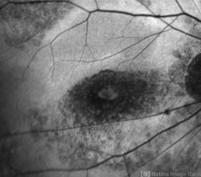
- Fundus autofluorescence image of the right eye in a highly myopic Caucasian patient who was screened for plaquenil toxicity elsewhere for 15 years. Medicine was stopped 3 years ago. She presented to us with deteriorationg central vision and scotoma for the past 3 years. FAF demonstrtaes classic symmetric bull's eye pattern of hypoautofluorescence in parafoveal area both eyes with some extension to the arcades, which is the result of severe plaquenil toxicity. Notice the hyperAF area surrounding the bull'e eye which is demonstarting stressed RPE cells filled with Lipofuscin. This area will likely turn to a larger hypoAF later. It is critical to diagnose it early to prevent the ongoing damage and loss of RPE and photoreceptors from the long term deposited medicine in RPE cells.


---thumb.jpg/image-square;max$79,0.ImageHandler "Bull's Eye Maculopathy")
---thumb.jpg/image-square;max$79,0.ImageHandler "Bull's Eye Maculopathy")


---thumb.jpg/image-square;max$79,0.ImageHandler "Chloroquine Toxicity")
---thumb.jpg/image-square;max$79,0.ImageHandler "Chloroquine Toxicity")