Search results (49 results)
-
 Vortex-pattern Exudative Retinal Detachment
Feb 22 2025 by CUI YUELING
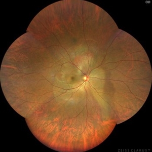
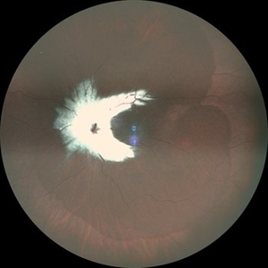
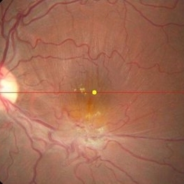
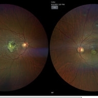
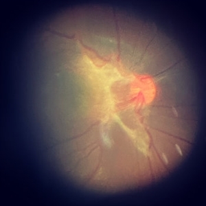
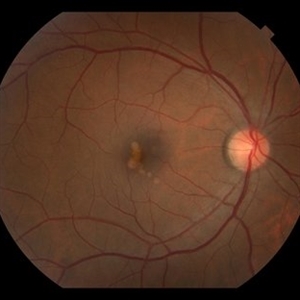
Patient: Male, 40 years old. Chief Complaint: Blurred vision and metamorphopsia in the left eye for more than 10 days. Past Medical History Hypertension for 4 years, with a highest recorded blood pressure of 160/80 mmHg. Currently controlled with oral "Nifedipine Sustained-Release Tablets, 2 tablets daily." Long-term history of heavy alcohol consumption and smoking. Ophthalmic Examination: Visual Acuity: Right eye (OD): 0.4 (uncorrected, no improvement with correction). Left eye (OS): 0.5 (-1.5DS = 1.0). Intraocular Pressure (IOP): OD: 15 mmHg. OS: 17 mmHg. Anterior Segment:Unremarkable. Fundus Examination: Right eye: Optic disc margins are clear, with a slightly reddish hue. Cup-to-disc ratio (C/D) = 0.2. A scalloped, orange-red elevated lesion is observed superior to the optic disc, with anterior displacement of the focal point. This is accompanied by a secondary, turbine-like exudative retinal detachment centered around the optic disc, involving the macula. The macular region shows scattered punctate yellow-white exudates. Diagnosis: Choroidal hemangioma with secondary exudative retinal detachment(OD).
Photographer: Cui yueling The First People's Hospital of Zunyi, Guizhou, Zunyi, China
Imaging device: Zeiss Clarus 500
Condition/keywords: choroidal hemangioma, exudative retinal detachment
-
 Photic Retinopathy
Jan 30 2025 by Juan Alberto Olivera Cueva
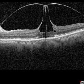
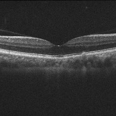
A 23-year-old male with a history of direct exposure to sunlight on several occasions, presenting blurred vision, comes for evaluation due to metamorphopsias of 3 months' evolution. The fundus photograph shows the presence of an epiretinal membrane. The OCT shows a hyperreflective line at the vitreomacular interface that causes traction to the layers of the inner retina, as well as distortion in the architecture of the macular region, with the presence of subfoveal detachment of the RPE.
Photographer: Dr. Juan Alberto Olivera Cueva, Escuela Militar de Medicina, Hospital Militar de Especialidades Oftalmológicas
Condition/keywords: epiretinal membrane (ERM)
-
 Photic Retinopathy
Jan 30 2025 by Juan Alberto Olivera Cueva
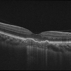
A 23 year-old male with a history of direct exposure to sunlight on several occasions, presenting blurred vision, comes for evaluation due to metamorphopsias of 3 months' evolution. The fundus photograph shows the presence of an epiretinal membrane. The OCT shows a hyperreflective line at the vitreomacular interface that causes traction to the layers of the inner retina, as well as distortion in the architecture of the macular region, with the presence of subfoveal detachment of the RPE.
Photographer: Dr. Juan Alberto Olivera Cueva, Escuela Militar de Medicina, Hospital Militar de Especialidades Oftalmológicas
Condition/keywords: MER, photic retinopathy
-
 Photic Retinopathy
Jan 29 2025 by Juan Alberto Olivera Cueva
A 23 year-old male with a history of direct exposure to sunlight on several occasions, presenting blurred vision, comes for evaluation due to metamorphopsias of 3 months' evolution. The fundus photograph shows the presence of an epiretinal membrane. The OCT shows a hyperreflective line at the vitreomacular interface that causes traction to the layers of the inner retina, as well as distortion in the architecture of the macular region, with the presence of subfoveal detachment of the RPE.
Photographer: Dr. Juan Alberto Olivera Cueva, Escuela Militar de Graduados de Sanidad, Hospital Militar de Especialidades Oftalmológico
Condition/keywords: Membrana Epirretiniana, MER, Photic retinopathy
-
 Photic Retinopathy
Jan 29 2025 by Juan Alberto Olivera Cueva
A 23-year-old male with a history of direct exposure to sunlight on several occasions, presenting blurred vision, comes for evaluation due to metamorphopsias of 3 months' evolution. The fundus photograph shows the presence of an epiretinal membrane. The OCT shows a hyperreflective line at the vitreomacular interface that causes traction to the layers of the inner retina, as well as distortion in the architecture of the macular region, with the presence of subfoveal detachment of the RPE.
Photographer: Dr. Juan Alberto Olivera Cueva, Escuela Militar de Graduados de Sanidad, Hospital Militar de Especialidades Oftalmológico
Condition/keywords: Membrana epirretiniana, MER, Photic Retinopathy
-
 Myelinated Nerve Fibres With Combined Hamartoma of Retina and RPE
Jul 31 2024 by Tejaswita Verma
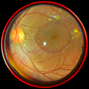
Fundus image of a 20 year old female who presented with metamorphopsia ,slightly blurred vision. BCVA was 6/9, epiretinal membrane present on central fundus examination with myelinated nerve fibres.
Photographer: DR. TEJASWITA VERMA
Imaging device: MIRANTE
Condition/keywords: combined hamartoma of retina and RPE, myelinated nerve fibers
-
 Best Disease
Apr 24 2024 by Marcelo Zas, MD PhD
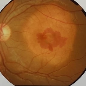
Best vitelliform macular dystrophy (BVMD) or Best disease. Is the most common autosomal dominant macular dystrophy. It involves the retinal pigment epithelium (RPE), and leads to a characteristic bilateral yellow “egg-yolk” appearance of the macula as you can see in this image. Essentially, BVMD is considered to have 6 clinical stages: Previtelliform, Vitelliform, Pseudohypopyon, Vitelleruptive, Atrophic and Choroidal neovascularization. As the disease progresses, patients may experience a slow, bilateral decrease in visual acuity, central scotoma, or metamorphopsia. With secondary CNV, visual decline can be rapid, however.
Photographer: Luciano Scorsetti MD
Condition/keywords: Macular Dystrophy
-
 Combined hamartoma of retina and retinal pigment epithelium
Aug 8 2023 by Navneet Mehrotra, DNB
A 20 year old female presented with decreased vision and metamorphopsia noticed in her left eye for one year. Other eye was normal. BCVA was 6/12 in her left eye.
Photographer: Dharti, Retina Care , Ahmedabad
Condition/keywords: Combined pigment epithelial and retinal hamartoma
-
 A rare case of a 45-year-old male with choroidal neovascular membrane in Familial Dominant Drusen (Doyne Honeycomb Drusen) in both eyes treated with intravitreal injections.
Nov 30 2022 by SHRADDHA ASHOK CHANDORKAR, DNB DO
A 45-year-old man presented with diminution of vision in both eyes with metamorphopsia, which was painless and gradually progressive in nature. BCVA at presentation were 6/40 and 6/36 for the right and left eye respectively. Anterior segment examination of both eyes was unremarkable. IOP were within normal limits. Fundus examination showed bilateral numerous yellowish white round closely spaced lesions extending radially from the vascular arcades till the periphery associated with an elevated grayish macular choroidal neovascular membrane (CNV) with multiple drusen in the macular area and posterior pole. Impression was Familial Dominant Drusen (Doyne Honeycomb Drusen) associated with CNVM, both eyes. Color fundus photograph and autofluorescence showed Familial Dominant Drusen with CNVM. Subsequently , the patient underwent periodic intravitreal injections of Ranibizumab in both eyes under guarded visual prognosis, for which he tolerated well.
Photographer: NATIONAL INSTITUTE OF OPHTHALMOLOGY, PUNE
Imaging device: ZEISS CLARUS
Condition/keywords: choroidal neovascular membrane (CNVM), Doyne's Honeycomb, FAMILIAL DOMINANT DRUSEN, IMIM (Online Mendelian Inheritance in Man), intravitreal injection, Malattia Leventinese
-
 Eiffel tower within the eye
Mar 31 2022 by Bhavani Sankaran, MS (Ophthalmology)
OCT of left eye of a 68 year old female patient who presented with complaints of metamorphopsia. Image depicts vitreomacular traction. BCVA OS 20/40.
Photographer: Dr. Bhavani Sankaran, MS, Aravind Eye Hospital, Madurai
Imaging device: Heidelberg Spectralis
Condition/keywords: vitreomacular interface disorders, vitreomacular traction (VMT)
-
 Epiretinal membrane
Nov 28 2021 by Jorge Berganza
22 yo female with metamorphopsia in the left eye.
Photographer: Jorge Berganza MD
Imaging device: Iphone XR
Condition/keywords: ERM, macular traction
-
 Postoperative OCT Showing Resolution of the Macular Fold
Jul 9 2021 by Anton Orlin, MD
This is an OCT demonstrating resolution of the macular fold following surgical repair. The anatomy of the macula has significantly improved, as have the patient’s symptoms of metamorphopsia.
Condition/keywords: macular fold
-
 Relentless Placoid Chorioretinitis
Jan 22 2021 by Renata Garcia Franco, Md
20-year-old male with reduction of vision in both eyes, scotoma and metamorphopsia. Widespread multiple chorioretinal lesions with RPE hyperplasia, which appear from posterior pole to peripheral retina and inactive choroidal neovascular membrane.
Photographer: Fatima Hernandez, Instituto de la Retina del Bajio SC
Imaging device: Zeiss
Condition/keywords: chorioretinitis
-
 Relentless Placoid Chorioretinitis
Jan 22 2021 by Renata Garcia Franco, Md
20-year-old male with reduction of vision in both eyes, scotoma and metamorphopsia. Widespread multiple chorioretinal lesions with RPE hyperplasia, which appear from posterior pole to peripheral retina.
Photographer: Fatima Hernandez, Instituto de la Retina del Bajio SC
Imaging device: Zeiss
Condition/keywords: chorioretinitis
-
 Circumscribed Choroidal Hemangioma
Jul 3 2020 by Dhaivat Shah
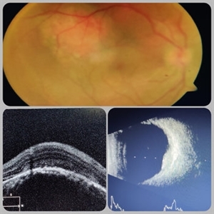
A 30-year-old young male presented with drop in vision in right eye since 1 year (6/60). Fundus examination revealed choroidal hemangioma superotemporal to macula. Choroidal hemangioma is an unusual benign vascular tumor of the choroid. It can be circumscribed solitary or diffuse tumor with the later having other systemic associations. Circumscribed choroidal hemangiomas (CCHs) are usually unilateral, unifocal hamartomatous vascular tumor affecting people in second to fourth decade. It appers as round to oval, orangish-red mass in posterior pole with smooth homogenous surface mostly present in macular and peripapillary area. Hyperopic shift is seen in sub-foveal tumors in contrast to para-foveal ones which are usually asymptomatic or present with metamorphopsia or photopsia and diminished vision secondary to exudative retinal detachment. B-scan shows highly reflective tumor without any shadowing or acoustic solidity with high anterior A scan spike. EDI-OCT here depicts a smooth gently sloping choridal mass with compressed choriocapillaries and enlarged medium and large choroidal vessels. Over a period of time structural abnormalities of the outer retina can be visualised. Ancillary testing using Fluorescein Angiography shows lacy hyper-fluorescence during early arterial phase followed by increased hyper-fluorescence due to progressive profuse leakage from pin point foci during arterial and venous phase. Indocyanine green angiography shows lacy diffuse fluorescent tumor in early phase followed by hypo-fluorescent tumor due to dye wash out in late phase. Intrinsic auto-fluorescence is also seen in CCHs from lipofuscin and fresh sub-retinal fluid. Tumor is relatively hyper-intense with respect to vitreous in T1-weighted images in iso-intense in T2-weighted images of MRI. Asymptomatic cases need no treatment, while patients showing vision loss with presence or absence of exudative retinal detachment can be treated with photodynamic therapy which is preferred treatment due to site specific action. Selective occlusion of choroidal neovascularization can be achieved while the neurosensory retinal layers and Bruch membrane are almost unaffected, leaving retinal function intact. Green or rarely red wavelength laser photocoagulation is used to create a chorioretinal adhesion and resolve the SRF. Other treatment modalities include Transpupilary thermotherapy, external beam irradiation, proton beam therapy, brachytherapy and gamma knife.
Photographer: Miss Deepika Nagle
Imaging device: Zeiss
Condition/keywords: B scan ultrasound, choroidal hemangioma, fundus photograph, optical coherence tomography (OCT), photodynamic therapy
-
 Choroidal Osteoma With Active CNVM
Apr 10 2020 by Dipak Nag, MBBS, FCPS, MSc, FRF
A 12-year-old boy visited our clinic for sudden, painless blurring of vision and metamorphopsia in left eye that he noticed 7 days back. His BCVA was 6/60 in left eye. Anterior segment examination was unremarkable. On fundus examination of his left eye showed a yellow-white lesion at the macula with well-defined geographic border and diffuse and mottled depigmentation of the overlying pigment epithelium, of which an elevated gray-green areas at the center with subretinal hemorrhage around. The right eye was found normal.
Photographer: Mr. Shamsuddin
Condition/keywords: choroidal neovascular membrane (CNVM)
-
 Spontaneous Resolution of Vitreomacular Traction
Dec 28 2019 by Anfisa Ayalon, MD
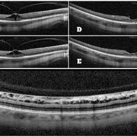
A 42-year-old highly myopic patient presented to the retina clinic with complains of metamorphopsia in his RE for the past months. SD-OCT revealed RE vitreomacular traction with stage 1 macular hole (A-B) and flat myopic retinoschisis (C). Visual acuity in the same eye was 6/30. After 3 weeks of follow up spontaneous resolution of vitreomacular traction was seen with full resolution of the symptoms. (D-E). Visual acuity remained without significant change.
Photographer: Anfisa Ayalon,MD., Meir Medical Center, Kfar Saba, Israel.
Condition/keywords: flat retinoschisis, macular hole, myopia, stage 1 macular hole, vitreomacular traction (VMT)
-
 Spontaneous Resolution of Vitreomacular Traction
Dec 28 2019 by Anfisa Ayalon, MD
A 42-year-old highly myopic patient presented to the retina clinic with complains of metamorphopsia for the past months. SD-OCT revealed RE vitreomacular traction with stage 1 macular hole (pictures from the left). After 3 weeks of follow up spontaneous resolution of vitreomacular traction (pictures from the right) was seen with full resolution of the symptoms.
Photographer: Anfisa Ayalon, MD., Meir Medical Center, Kfar Saba, Israel.
Condition/keywords: macular hole, myopia, spontaneous resolution, stage 1 macular hole, vitreomacular traction (VMT)
-
 Unilateral PIC Following Recent Influenza Vaccine
Jan 6 2019 by John S. King, MD
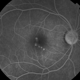
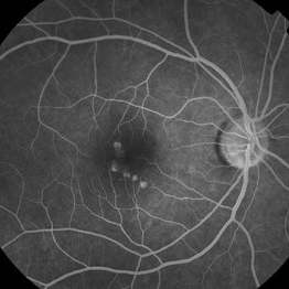
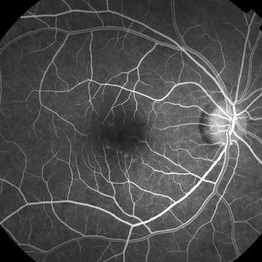
FA 2 minute 42-year-old African American female with high myopia and type 2 diabetes, presented to her eye doctor with distortion in the right eye "that looked like seeing through a coke bottle". She denied any photopsias or other symptoms. She received an influenza vaccine two weeks before onset of metamorphopsia. I saw her about a month after symptoms began. Va cc 20/30 OD J1 (20/15 J1+ OS); a/c and vitreous without cell or flare. Posterior pole OD showed yellowish, rounded small to medium RPE pigment alterations without heme or exudes (OS U/R). FA showed early focal areas of hyperfluorescence that stained in the later frames without CNVM or CME; rare MA inferiorly. The OCT showed some focal PEDs with some possible overlying SRHRM. We discussed options and decided to try a medrol dose pack. A few weeks later she was 20/20 J1, with minimal to no symptoms; OCT shows near total resolution of PEDs.
Photographer: Kay Dalby
Imaging device: Topcon 50
Condition/keywords: punctate inner choroidopathy (PIC), white dot syndrome
-
 Unilateral PIC Following Recent Influenza Vaccine
Jan 6 2019 by John S. King, MD
FA 5 minute 42-year-old African American female with high myopia and type 2 diabetes, presented to her eye doctor with distortion in the right eye "that looked like seeing through a Coke bottle." She denied any photopsias or other symptoms. She received an influenza vaccine two weeks before onset of metamorphopsia. I saw her about a month after symptoms began. Va cc 20/30 OD J1 (20/15 J1+ OS); a/c and vitreous without cell or flare. Posterior pole OD showed yellowish, rounded small to medium RPE pigment alterations without heme or exudes (OS U/R). FA showed early focal areas of hyperfluorescence that stained in the later frames without CNVM or CME; rare MA inferiorly. The OCT showed some focal PEDs with some possible overlying SRHRM. We discussed options and decided to try a medrol dose pack. A few weeks later she was 20/20 J1, with minimal to no symptoms; OCT shows near total resolution of PEDs.
Photographer: Kay Dalby
Imaging device: Topcon 50
Condition/keywords: punctate inner choroidopathy (PIC), white dot syndrome
-
 Unilateral PIC following recent influenza vaccine
Jan 6 2019 by John S. King, MD
FA 22 seconds 42-year-old African American female with high myopia and type 2 diabetes, presented to her eye doctor with distortion in the right eye "that looked like seeing through a Coke bottle." She denied any photopsias or other symptoms. She received an influenza vaccine two weeks before onset of metamorphopsia. I saw her about a month after symptoms began. Va cc 20/30 OD J1 (20/15 J1+ OS); a/c and vitreous without cell or flare. Posterior pole OD showed yellowish, rounded small to medium RPE pigment alterations without heme or exudes (OS U/R). FA showed early focal areas of hyperfluorescence that stained in the later frames without CNVM or CME; rare MA inferiorly. The OCT showed some focal PEDs with some possible overlying SRHRM. We discussed options and decided to try a medrol dose pack. A few weeks later she was 20/20 J1, with minimal to no symptoms; OCT shows near total resolution of PEDs.
Photographer: Kay Dalby
Imaging device: Topcon 50
Condition/keywords: punctate inner choroidopathy (PIC), white dot syndrome
-
 Unilateral PIC Following Recent Influenza Vaccine
Jan 6 2019 by John S. King, MD
42-year-old African American female with high myopia and type 2 diabetes, presented to her eye doctor with distortion in the right eye "that looked like seeing through a Coke bottle." She denied any photopsias or other symptoms. She received an influenza vaccine two weeks before onset of metamorphopsia. I saw her about a month after symptoms began. Va cc 20/30 OD J1 (20/15 J1+ OS); a/c and vitreous without cell or flare. Posterior pole OD showed yellowish, rounded small to medium RPE pigment alterations without heme or exudes (OS U/R). FA showed early focal areas of hyperfluorescence that stained in the later frames without CNVM or CME; rare MA inferiorly. The OCT showed some focal PEDs with some possible overlying SRHRM. We discussed options and decided to try a medrol dose pack. A few weeks later she was 20/20 J1, with minimal to no symptoms; OCT shows near total resolution of PEDs.
Photographer: Kay Dalby
Imaging device: Topcon 50
Condition/keywords: punctate inner choroidopathy (PIC), white dot syndrome
-
 Unilateral PIC Following Recent Influenza Vaccine
Jan 6 2019 by John S. King, MD
OCT few weeks after initial visit. 42-year-old African American female with high myopia and type 2 diabetes, presented to her eye doctor with distortion in the right eye "that looked like seeing through a Coke bottle." She denied any photopsias or other symptoms. She received an influenza vaccine two weeks before onset of metamorphopsia. I saw her about a month after symptoms began. Va cc 20/30 OD J1 (20/15 J1+ OS); a/c and vitreous without cell or flare. Posterior pole OD showed yellowish, rounded small to medium RPE pigment alterations without heme or exudes (OS U/R). FA showed early focal areas of hyperfluorescence that stained in the later frames without CNVM or CME; rare MA inferiorly. The OCT showed some focal PEDs with some possible overlying SRHRM. We discussed options and decided to try a medrol dose pack. A few weeks later she was 20/20 J1, with minimal to no symptoms; OCT shows near total resolution of PEDs.
Photographer: Kay Dalby
Imaging device: Topcon 50
Condition/keywords: punctate inner choroidopathy (PIC), white dot syndrome
-
 Unilateral PIC Following Recent Influenza Vaccine
Jan 6 2019 by John S. King, MD
Initial OCT. 42-year-old African American female with high myopia and type 2 diabetes, presented to her eye doctor with distortion in the right eye "that looked like seeing through a Coke bottle." She denied any photopsias or other symptoms. She received an influenza vaccine two weeks before onset of metamorphopsia. I saw her about a month after symptoms began. Va cc 20/30 OD J1 (20/15 J1+ OS); a/c and vitreous without cell or flare. Posterior pole OD showed yellowish, rounded small to medium RPE pigment alterations without heme or exudes (OS U/R). FA showed early focal areas of hyperfluorescence that stained in the later frames without CNVM or CME; rare MA inferiorly. The OCT showed some focal PEDs with some possible overlying SRHRM. We discussed options and decided to try a medrol dose pack. A few weeks later she was 20/20 J1, with minimal to no symptoms; OCT shows near total resolution of PEDs.
Photographer: Kay Dalby
Imaging device: Topcon 50
Condition/keywords: punctate inner choroidopathy (PIC), white dot syndrome
-
 Central Serous Chorioretinopathy : Smartphone Fundus Image
Dec 14 2018 by Prithvi Chandrakanth
A 42-year-old male with diminution of vision in the left eye since one week, uncorrected visual acuity was 6/18 improving with hyperopic lens to 6/9. H/o Metamorphopsia was present.
Photographer: Dr.Prithvi Chandrakanth, Dr.Chandrakanth Malabar Nethralaya, Kozhikode.
Imaging device: Trash To Treasure (T3) Retcam - Smartphone Fundus Camera
Condition/keywords: central serous chorioretinopathy (CSCR), smartphone fundus photography
 Loading…