 Initializing download.
-
 By CUI YUELING
Co-author(s): Tan wei?Hua hengyan?Song zhaoxi?Fulili?Wang tao - Uploaded on Feb 22, 2025.
- Last modified by CUI YUELING on Feb 28, 2025.
- Rating
- Appears in
- Miscellaneous
- Condition/keywords
- exudative retinal detachment, choroidal hemangioma
- Photographer
- Cui yueling The First People's Hospital of Zunyi, Guizhou, Zunyi, China
- Imaging device
-
Scanning laser ophthalmoscope
Zeiss Clarus 500 - Description
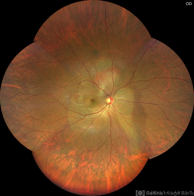
- Patient: Male, 40 years old. Chief Complaint: Blurred vision and metamorphopsia in the left eye for more than 10 days. Past Medical History Hypertension for 4 years, with a highest recorded blood pressure of 160/80 mmHg. Currently controlled with oral "Nifedipine Sustained-Release Tablets, 2 tablets daily." Long-term history of heavy alcohol consumption and smoking. Ophthalmic Examination: Visual Acuity: Right eye (OD): 0.4 (uncorrected, no improvement with correction). Left eye (OS): 0.5 (-1.5DS = 1.0). Intraocular Pressure (IOP): OD: 15 mmHg. OS: 17 mmHg. Anterior Segment:Unremarkable. Fundus Examination: Right eye: Optic disc margins are clear, with a slightly reddish hue. Cup-to-disc ratio (C/D) = 0.2. A scalloped, orange-red elevated lesion is observed superior to the optic disc, with anterior displacement of the focal point. This is accompanied by a secondary, turbine-like exudative retinal detachment centered around the optic disc, involving the macula. The macular region shows scattered punctate yellow-white exudates. Diagnosis: Choroidal hemangioma with secondary exudative retinal detachment(OD).


---thumb.jpg/image-square;max$79,0.ImageHandler "Harada's with Exudative RD")
---thumb.jpg/image-square;max$79,0.ImageHandler "Harada's with Exudative RD")


---thumb.jpg/image-square;max$79,0.ImageHandler "vitreous snowballs, peripheral retinal neovascularization, inferior snowbanking, vascular sheathing, and peripheral exudative retinal detachment")
---thumb.jpg/image-square;max$79,0.ImageHandler "vitreous haze and retinal detachment")