 Initializing download.
-
 By Dipak Nag, MBBS, FCPS, MSc, FRF
Govt.
Co-author(s): Dr Rinku Paul - Uploaded on Apr 10, 2020.
- Last modified by Caroline Bozell on Apr 10, 2020.
- Rating
- Appears in
- 10-Apr-2020
- Condition/keywords
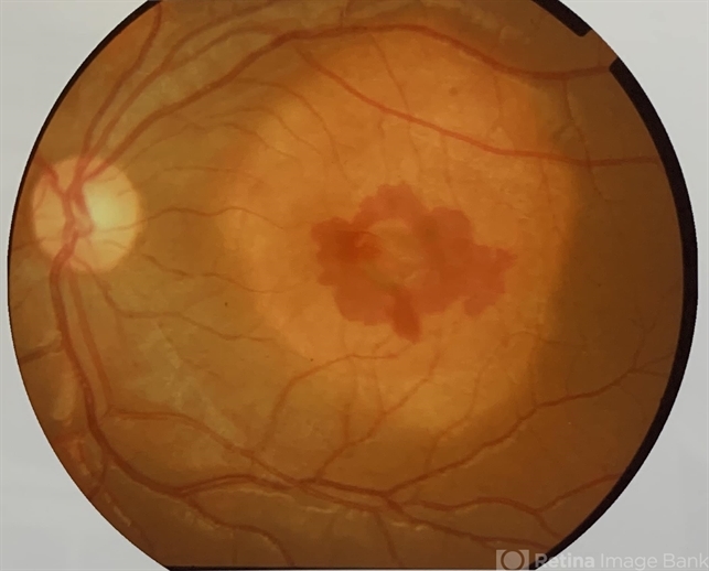
- choroidal neovascular membrane (CNVM)
- Photographer
- Mr. Shamsuddin
- Imaging device
- Fundus camera
- Description
- A 12-year-old boy visited our clinic for sudden, painless blurring of vision and metamorphopsia in left eye that he noticed 7 days back. His BCVA was 6/60 in left eye. Anterior segment examination was unremarkable. On fundus examination of his left eye showed a yellow-white lesion at the macula with well-defined geographic border and diffuse and mottled depigmentation of the overlying pigment epithelium, of which an elevated gray-green areas at the center with subretinal hemorrhage around. The right eye was found normal.


