Search results (49 results)
-
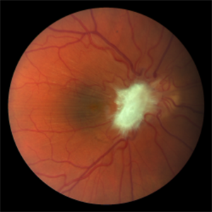
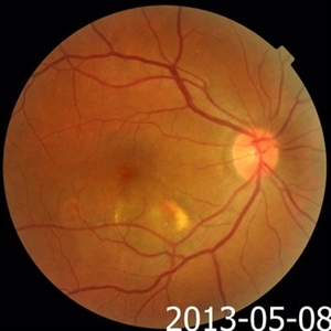
 Pigment Epithelial Detachment late FA with small occult CNV
Jul 6 2012 by Tarek S. Hassan, MD, FASRS
72-year-old man with VA loss and metamorphopsia of 2 months duration. PED found, testing done to rule out CNV. Very suspicious for CNV in superonasal fovea/parafovea.
Condition/keywords: choroidal neovascularization (CNV), pigment epithelial detachment (PED)
-
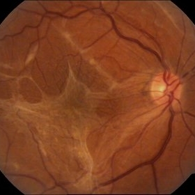 Epiretinal membrane - Fundus photograph
Feb 5 2014 by Gerardo Garcia-Aguirre, MD
Fundus photograph of a 62 year old female with metamorphopsia and decreased visual acuity. A stage 2 epiretinal membrane is observed, causing distortion of the retinal vasculature.
Photographer: Gerardo Garcia-Aguirre, MD
Condition/keywords: epiretinal membrane (ERM)
-
 Pigment Epithelial Detachment
Jul 6 2012 by Tarek S. Hassan, MD, FASRS
72-year-old man with VA loss and metamorphopsia of 2 months duration. PED found, testing done to rule out CNV.
Condition/keywords: metamorphopsia, pigment epithelial detachment (PED)
-
 Myopic Choroidal Neovascularization
Aug 23 2012 by Gabriela Lopezcarasa Hernandez, MD
19-year-old male who complains of scotoma and metamorphopsias.
Photographer: Gabriela Lopezcarasa Hernandez, Macular Retina Consultores
Imaging device: Heidelberg Spectralis
Condition/keywords: choroidal neovascularization (CNV), myopia
-
 Type 1A Macular Telangiectasia - Fundus photograph
Nov 11 2013 by Gerardo Garcia-Aguirre, MD
Fundus photograph of a 43-year-old male complaining of mild metamorphopsia in OS. BCVA 20/25. Some hard exudates and telangiectatic vessels are observed inferior and temporal to the fovea.
Condition/keywords: macular telangiectasia
-
 Central Serous Chorioretinopathy : Smartphone Fundus Image
Dec 14 2018 by Prithvi Chandrakanth
A 42-year-old male with diminution of vision in the left eye since one week, uncorrected visual acuity was 6/18 improving with hyperopic lens to 6/9. H/o Metamorphopsia was present.
Photographer: Dr.Prithvi Chandrakanth, Dr.Chandrakanth Malabar Nethralaya, Kozhikode.
Imaging device: Trash To Treasure (T3) Retcam - Smartphone Fundus Camera
Condition/keywords: central serous chorioretinopathy (CSCR), smartphone fundus photography
-
 Spontaneous Macular Hemorrhage in High Myopia
Jul 7 2015 by Hamid Ahmadieh, MD
A highly myopic 30-year-old-man noticed sudden drop of vision and metamorphopsia in his left eye. Color fundus photograph showed a macular haemorrhage (Fig. a) which partially resolved spontaneously after 6 weeks ( Fig. b). Four months later, VA improved to 20/40 and the image distortion markedly resolved ( Fig. c). Lacquer cracks were visible.
Photographer: Soulmaz Shahmohammad, Negah Eye Center, Tehran, Iran
Condition/keywords: color fundus photograph, high myopia, macular hemorrhage
-
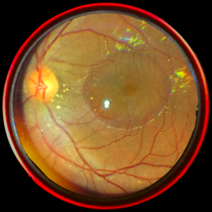
---thumb.jpg/image-square;max$300,300.ImageHandler) Polypoidal Choroidal Vasculopathy
Polypoidal Choroidal Vasculopathy
Jul 13 2013 by Hamid Ahmadieh, MD
Late phase FA and ICG images of the right eye of a 55-year-old woman with decreased vision and metamorphopsia due to PCV.
Photographer: Elham Salehi, Negah Eye Center, Tehran
Imaging device: Heidelberg Spectralis
Condition/keywords: indocyanine green (ICG) angiography, polypoidal choroidal vasculopathy (PCV)
-
 Pre-Retinal Fibrosis Following Endogenous Fungal Endophthalmitis
Aug 23 2018 by Matthew Dombrow, MD
20-year-old male presents with posterior uveitis with a chain of white infiltrates stemming from the optic nerve. While treated, his infiltrates retracted and hemorrhage occured. Neovascularization of the disc developed and underwent Avastin treatment in addition to his oral anti-fungals and intravitreal anti-fungals. Patient was lost to follow up and presented with severe pre-retinal fibrosis 6 years later. Acuity is 20/40-1 with significant metamorphopsia.
Photographer: Patricia Candrea, COA, Connecticut Retina Consultants, LLC
Imaging device: Canon
Condition/keywords: pre-retinal membrane
-
---thumb.jpg/image-square;max$300,300.ImageHandler) Polypoidal Choroidal Vasculopathy
Polypoidal Choroidal Vasculopathy
Jul 13 2013 by Hamid Ahmadieh, MD
FAF image of the right eye of a 55-year-old woman with decreased vision and metamorphopsia due to PCV.
Photographer: Elham Salehi, Negah Eye Center, Tehran
Imaging device: Heidelberg Spectralis
Condition/keywords: fundus autofluorescence (FAF), polypoidal choroidal vasculopathy (PCV)
-
 Polypoidal Choroidal Vasculopathy
Jul 13 2013 by Hamid Ahmadieh, MD
FA and ICG images of the right eye of a 55-year-old woman with decreased vision and metamorphopsia due to PCV.
Photographer: Elham Salehi, Negah Eye Center, Tehran
Imaging device: Heidelberg Spectralis
Condition/keywords: indocyanine green (ICG) angiography, polypoidal choroidal vasculopathy (PCV)
-
---thumb.png/image-square;max$300,300.ImageHandler) Inflammatory CNV- PIC
Inflammatory CNV- PIC
Mar 19 2014 by Roy Schwartz, MD
RE color picture of a 21-year-old girl with mild myopia -1.5 admitted with RE metamorphopsiae and blurred vision. A RE CNV was seen with BE leakage points picture taken before RE Bevacizumab injection.
Photographer: galit yair pur
Condition/keywords: choroidal neovascularization (CNV), inflammatory choroidopathy, punctate inner choroidopathy (PIC)
-
---thumb.jpg/image-square;max$300,300.ImageHandler) Inflammatory CNV- PIC
Inflammatory CNV- PIC
Mar 20 2014 by Roy Schwartz, MD
RE OCT of a 21-year-old girl with mild myopia -1.5 admitted with RE metamorphopsiae and blurred vision. A RE CNV was seen with BE leakage points picture shows a hyperreflective mass- CNV after RE Bevacizumab injection.
Photographer: Galit Yair Pur
Condition/keywords: choroidal neovascularization (CNV), inflammatory choroidopathy, punctate inner choroidopathy (PIC)
-
 Polypoidal Choroidal Vasculopathy
Jul 13 2013 by Hamid Ahmadieh, MD
Color fundus photograph of the right eye of a 55-year-old woman with decreased vision and metamorphopsia due to PCV.
Photographer: Elham Salehi, Negah Eye Center, Tehran
Imaging device: Topcon Fundus Camera
Condition/keywords: polypoidal choroidal vasculopathy (PCV)
-
 Retinal Angiomatous Proliferation
Sep 10 2018 by Gabriela Lopezcarasa Hernandez, MD
75-year-old patient with decrease in visual acuity right eye with metamorphopsia, in the FA and ICG we can see a RAP lesion.
Photographer: Azucena Rios
Imaging device: Heidelberg Spectralis
Condition/keywords: FA mid phase, indocyanine green (ICG) angiography, RAP lesion, retinal angiomatous proliferation (RAP)
-
---thumb.jpg/image-square;max$300,300.ImageHandler) Inflammatory CNV- PIC
Inflammatory CNV- PIC
Mar 20 2014 by Roy Schwartz, MD
RE OCT of a 21-year-old girl with mild myopia -1.5 admitted with RE metamorphopsiae and blurred vision. A RE CNV was seen with BE leakage points on FA. Picture shows a hyperreflective mass- CNV before RE Bevacizumab injection.
Photographer: Galit Yair Pur
Condition/keywords: choroidal neovascularization (CNV), inflammatory choroidopathy, punctate inner choroidopathy (PIC)
-
---thumb.jpg/image-square;max$300,300.ImageHandler) Inflammatory CNV- PIC
Inflammatory CNV- PIC
Mar 19 2014 by Roy Schwartz, MD
RE FA of a 21-year-old girl with mild myopia -1.5 admitted with RE metamorphopsiae and blurred vision. A RE CNV was seen with BE leakage points picture shows the leakage from the CNV and hyperfluorescent dots.
Photographer: Galit Yair Pur
Condition/keywords: choroidal neovascularization (CNV), inflammatory choroidopathy, punctate inner choroidopathy (PIC)
-
 Spontaneous Macular Hemorrhage in High Myopia
Jul 7 2015 by Hamid Ahmadieh, MD
A highly myopic 30-year-old-man noticed sudden drop of vision and metamorphopsia in his left eye. OCT showed a macular haemorrhage (Fig. a) which partially resolved spontaneously after 6 weeks ( Fig. b). Four months later, VA improved to 20/40 and the image distortion markedly resolved ( Fig. c).
Photographer: Soulmaz Shahmohammad, Negah Eye Center, Tehran, Iran
Imaging device: Specteralis
Condition/keywords: high myopia, macular hemorrhage, optical coherence tomography (OCT)
-
---thumb.JPG/image-square;max$300,300.ImageHandler) Inflammatory CNV- PIC
Inflammatory CNV- PIC
Mar 19 2014 by Roy Schwartz, MD
RE color picture of a 21-year-old girl with mild myopia -1.5 admitted with RE metamorphopsiae and blurred vision. A RE CNV was seen with BE leakage points on FA. Picture was taken before RE Bevacizumab injection.
Photographer: Galit Yair Pur
Condition/keywords: choroidal neovascularization (CNV), inflammatory choroidopathy, punctate inner choroidopathy (PIC)
-
---thumb.jpg/image-square;max$300,300.ImageHandler) Inflammatory CNV- PIC
Inflammatory CNV- PIC
Mar 19 2014 by Roy Schwartz, MD
LE FA of a 21-year-old girl with mild myopia -1.5 admitted with RE metamorphopsiae and blurred vision. A RE CNV was seen with BE leakage points.
Photographer: Galit Yair Pur
Condition/keywords: choroidal neovascularization (CNV), inflammatory choroidopathy, punctate inner choroidopathy (PIC)
-
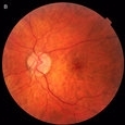
---thumb.jpg/image-square;max$300,300.ImageHandler) Metamorphopsia
Metamorphopsia
Oct 15 2013 by Maurice F. Rabb
A 38 year old lady presented with metamorphopsia in the left eye of two days' duration. Vision was 20/30 OD and 20/40 OS. Pigmentary changes were seen in the posterior pole consistent with pattern dystrophy. A year later she had acute loss of vision in the right eye to 20/100 but no subretinal neovascularization was evident. Subsequently this recovered to the level of 20/50.
Condition/keywords: metamorphopsia
-
 Relentless Placoid Chorioretinitis
Jan 22 2021 by Renata Garcia Franco, Md
20-year-old male with reduction of vision in both eyes, scotoma and metamorphopsia. Widespread multiple chorioretinal lesions with RPE hyperplasia, which appear from posterior pole to peripheral retina and inactive choroidal neovascular membrane.
Photographer: Fatima Hernandez, Instituto de la Retina del Bajio SC
Imaging device: Zeiss
Condition/keywords: chorioretinitis
-
---thumb.jpg/image-square;max$300,300.ImageHandler) Metamorphopsia
Metamorphopsia
Oct 15 2013 by Maurice F. Rabb
A 38 year old lady presented with metamorphopsia in the left eye of two days' duration. Vision was 20/30 OD and 20/40 OS. Pigmentary changes were seen in the posterior pole consistent with pattern dystrophy. A year later she had acute loss of vision in the right eye to 20/100 but no subretinal neovascularization was evident. Subsequently this recovered to the level of 20/50.
Condition/keywords: metamorphopsia
-
 Relentless Placoid Chorioretinitis
Jan 22 2021 by Renata Garcia Franco, Md
20-year-old male with reduction of vision in both eyes, scotoma and metamorphopsia. Widespread multiple chorioretinal lesions with RPE hyperplasia, which appear from posterior pole to peripheral retina.
Photographer: Fatima Hernandez, Instituto de la Retina del Bajio SC
Imaging device: Zeiss
Condition/keywords: chorioretinitis
 Loading…