 Initializing download.
-
 By Dhaivat Shah
Sankara Nethralaya
Co-author(s): Dr Amit Porwal, Dr Rahul Singh - Uploaded on Jul 3, 2020.
- Last modified by Caroline Bozell on Jul 7, 2020.
- Rating
- Appears in
- Imaging marvels
- Condition/keywords
- choroidal hemangioma, optical coherence tomography (OCT), fundus photograph, B scan ultrasound, photodynamic therapy
- Photographer
- Miss Deepika Nagle
- Imaging device
-
Fundus camera
Zeiss - Description
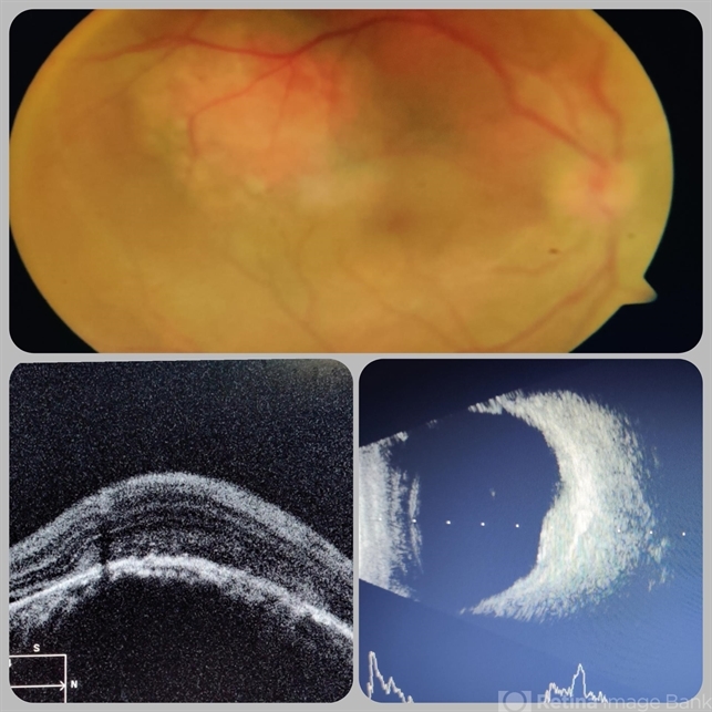
- A 30-year-old young male presented with drop in vision in right eye since 1 year (6/60). Fundus examination revealed choroidal hemangioma superotemporal to macula. Choroidal hemangioma is an unusual benign vascular tumor of the choroid. It can be circumscribed solitary or diffuse tumor with the later having other systemic associations. Circumscribed choroidal hemangiomas (CCHs) are usually unilateral, unifocal hamartomatous vascular tumor affecting people in second to fourth decade. It appers as round to oval, orangish-red mass in posterior pole with smooth homogenous surface mostly present in macular and peripapillary area. Hyperopic shift is seen in sub-foveal tumors in contrast to para-foveal ones which are usually asymptomatic or present with metamorphopsia or photopsia and diminished vision secondary to exudative retinal detachment. B-scan shows highly reflective tumor without any shadowing or acoustic solidity with high anterior A scan spike. EDI-OCT here depicts a smooth gently sloping choridal mass with compressed choriocapillaries and enlarged medium and large choroidal vessels. Over a period of time structural abnormalities of the outer retina can be visualised. Ancillary testing using Fluorescein Angiography shows lacy hyper-fluorescence during early arterial phase followed by increased hyper-fluorescence due to progressive profuse leakage from pin point foci during arterial and venous phase. Indocyanine green angiography shows lacy diffuse fluorescent tumor in early phase followed by hypo-fluorescent tumor due to dye wash out in late phase. Intrinsic auto-fluorescence is also seen in CCHs from lipofuscin and fresh sub-retinal fluid. Tumor is relatively hyper-intense with respect to vitreous in T1-weighted images in iso-intense in T2-weighted images of MRI. Asymptomatic cases need no treatment, while patients showing vision loss with presence or absence of exudative retinal detachment can be treated with photodynamic therapy which is preferred treatment due to site specific action. Selective occlusion of choroidal neovascularization can be achieved while the neurosensory retinal layers and Bruch membrane are almost unaffected, leaving retinal function intact. Green or rarely red wavelength laser photocoagulation is used to create a chorioretinal adhesion and resolve the SRF. Other treatment modalities include Transpupilary thermotherapy, external beam irradiation, proton beam therapy, brachytherapy and gamma knife.

")


")
")
")
")
")
")